1B · The Cell Membrane
A cellular skin of sorts wraps the outside of the cell. This is the cell membrane, also called the plasma membrane. The nucleus sits in the middle holding the cell’s DNA and functions something like the cell’s brain. The other organelles work a bit like organs work inside us, each carrying out a different function, together covering everything a cell needs.
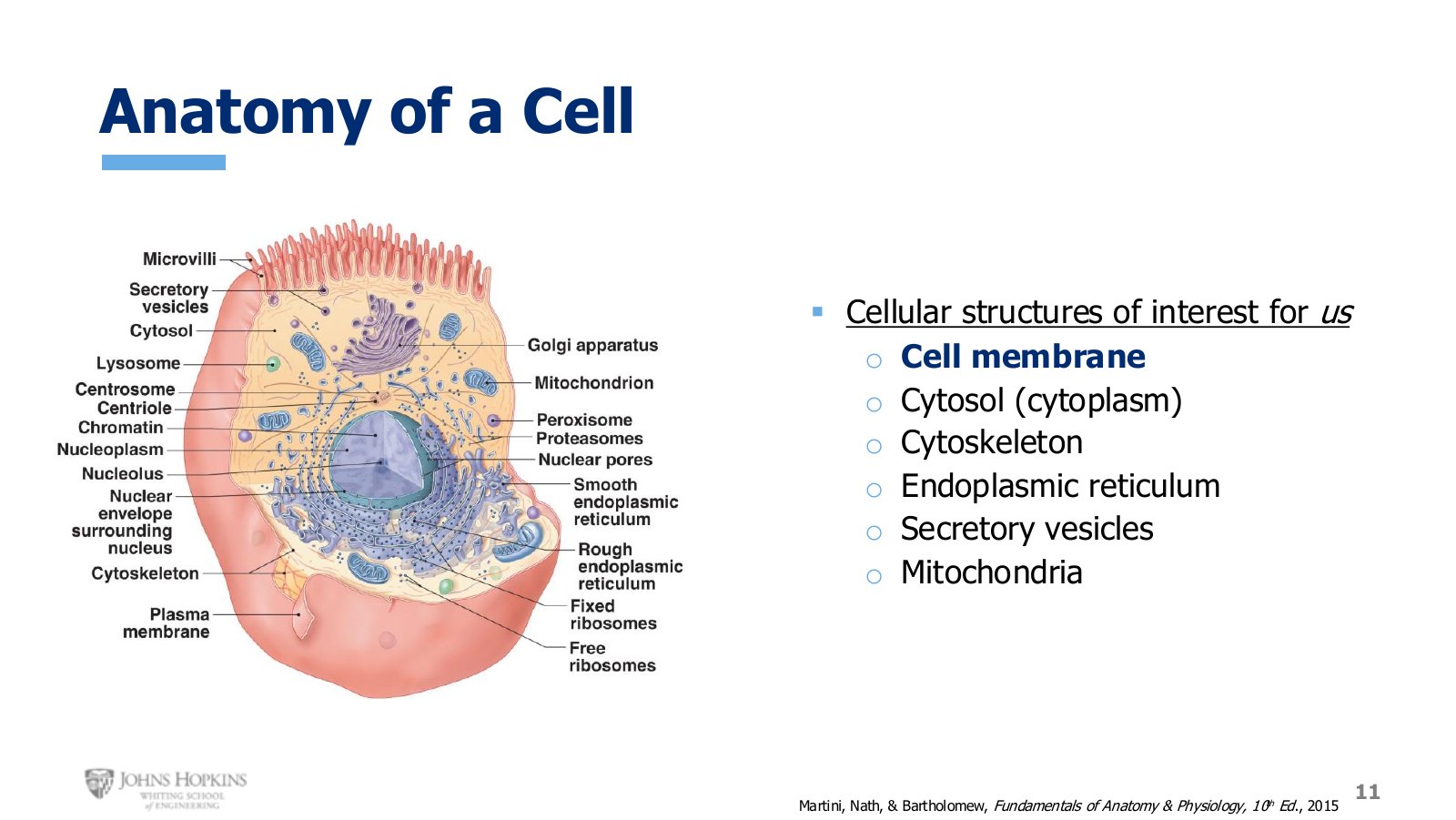
Slide 11. Basic cell anatomy. Six structures on this diagram recur all semester.
Six structures matter for this course.
Cell membrane. The organelle referenced most this semester.
Cytosol, also called cytoplasm. The fluid environment inside the cell in which everything else is dissolved or suspended.
Cytoskeleton. A network of structural proteins giving the cell shape and form. Important for muscle tissue.
Endoplasmic reticulum. A storage organelle. In muscle tissue it stores the ions critical for contraction.
Secretory vesicles. What neural cells use to release the chemical messengers that tell the rest of the body what to do.
Mitochondria. Energy production, touched on briefly for muscle tissue.
Four purposes of the membrane
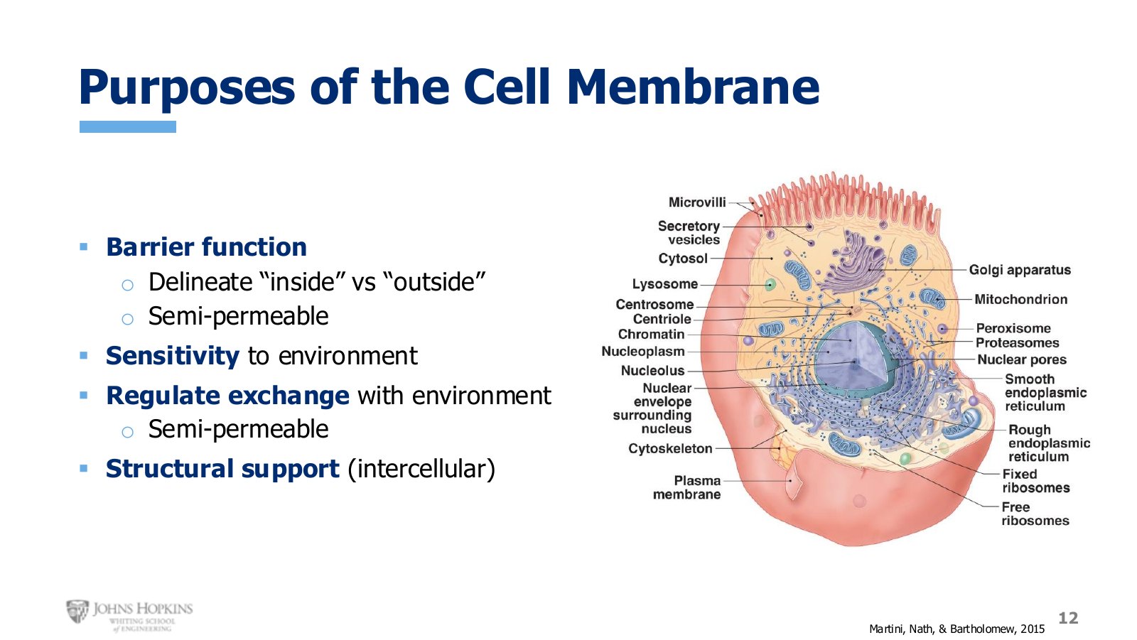
Slide 12. The four jobs the membrane performs. Everything in the rest of this lecture serves one of them.
Barrier function. It physically isolates the inside of the cell from everything outside. The barrier is not impermeable, though. It is semi-permeable, meaning selective about what gets through.
Sensitivity to the environment. It lets the cell receive chemical messages from cells and tissues both nearby and far away.
Regulated exchange. Structures embedded in the membrane control which molecules cross and when.
Structural support. Embedded molecules bind the membrane to nearby tissues and other cell membranes, anchoring adjacent cells physically to each other.
Fluid compartments
The body is about 60% water, varying with age and sex. More precisely, the body is a fluid solution: water plus non-water molecules, with the water dissolving most of them.
Solute: anything that dissolves in a liquid. Its ability to do so is its solubility.
Solvent: the liquid doing the dissolving. In the human body, water.
Solution: solvent plus solute.
Concentration: the ratio of solute amount to volume of solution.
Body solutes include sodium, potassium, calcium, glucose, hormones, proteins, amino acids, waste products, and gases.
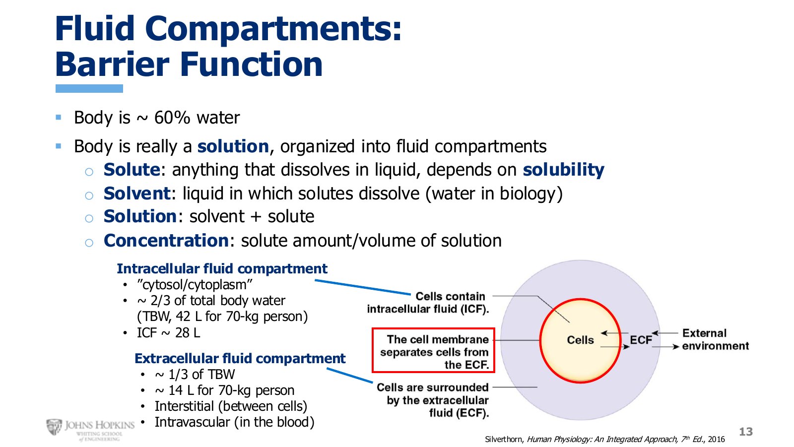
Slide 13. Body water divided into compartments. The cell membrane is the wall between the two major ones.
All water inside cells is collectively intracellular fluid. All water outside cells is extracellular fluid. The cell membrane physically divides them.
The intracellular fluid compartment is really just the cytoplasm of every cell in the body. We treat that aggregate as one compartment because it is remarkably uniform from cell to cell. The extracellular fluid compartment is further divided into the interstitial fluid found between cells and the intravascular fluid found in blood vessels, which is plasma, the watery portion of blood.
Compartment volumes, 70 kg person
Compartment | Fraction of TBW | Volume |
|---|---|---|
Total body water (60% of 70 kg) | — | 42 L |
Intracellular fluid | 2/3 | 28 L |
Extracellular fluid | 1/3 | 14 L |
Phospholipids and the barrier
The membrane is described as a phospholipid bilayer. The phospholipids are what make it such a good barrier between the watery intracellular and extracellular compartments.
The name is descriptive: part of the molecule is lipid, part contains phosphorus. A phospholipid has two fatty acid carbon chains at the bottom, the fatty acid tails, bound to a glycerol backbone, above which sits a phosphate group, phosphorus surrounded by four oxygens.
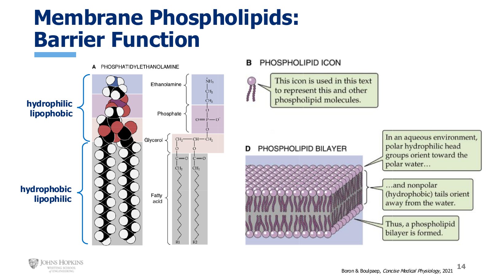
Slide 14. A phospholipid drawn two ways, then the stick-figure shorthand, then the bilayer they form.
If you have had vinegar salad dressing, you know oil and water do not get along. The fatty acid tails share that aversion, so that end of the molecule is hydrophobic (water-fearing) and lipophilic (fat-loving). The glycerol and phosphate dissolve easily in water and struggle to interact with fat, so that end is hydrophilic and lipophobic.
Submerge phospholipids in the watery environment of the body and the best way for them to form a membrane while still interacting with water the way they want is a double layer. Hydrophilic phosphate heads face outward toward the watery compartments. Hydrophobic tails face inward toward each other, avoiding water as much as they can, and in doing so create a strongly hydrophobic core in the middle of the membrane.
The watery compartments on either side cannot pass through and mix, because water cannot penetrate that lipid core. That is the whole barrier mechanism.
Membrane proteins
The membrane is only about 50% lipid by volume. The other half is protein. Membrane proteins are what deliver sensitivity, exchange, and structural support.
Proteins are either integral, physically embedded in the membrane and often though not always spanning the entire bilayer, or peripheral, attached to an integral protein but not bound to the membrane itself.
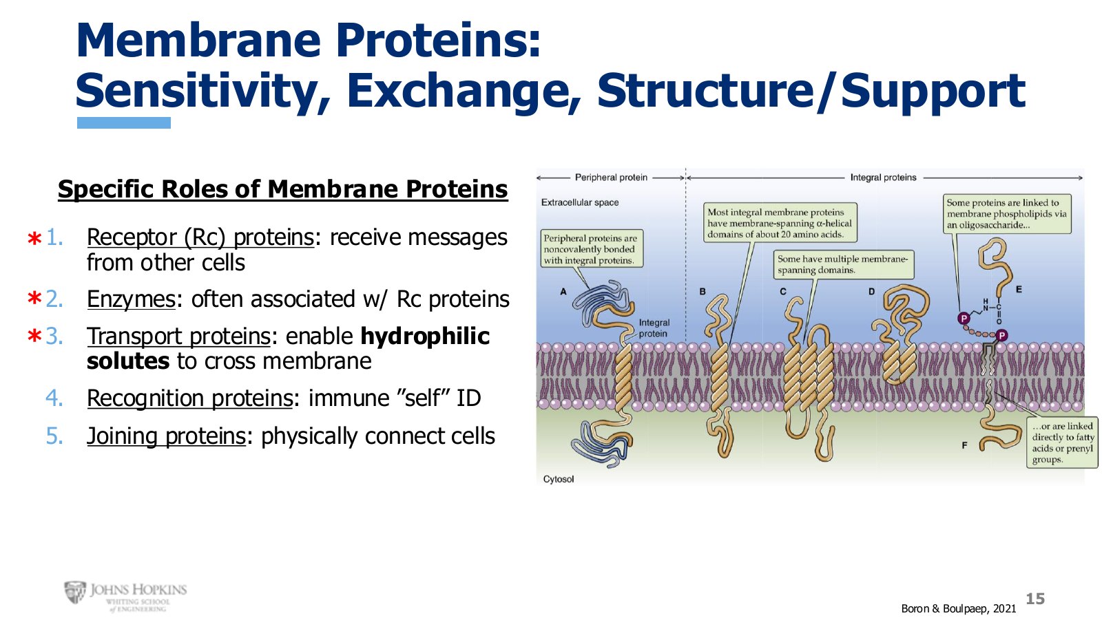
Slide 15. The five roles of membrane proteins. Receptors, enzymes, and transporters are the three this course pursues.
Receptors receive and translate chemical messages from outside the cell.
Enzymes catalyze chemical reactions, often though not always working alongside membrane receptors.
Transporters escort specific solutes through the membrane at the appropriate time.
Recognition proteins identify the cell to the immune system as native rather than foreign. Not covered in this course.
Joining proteins anchor a cell to its underlying connective tissue base or to nearby cells. Covered only briefly, under cardiac muscle structure.
Ligands and receptor types
A ligand is any chemical that binds to a membrane receptor. It could be a hormone like insulin or thyroid hormone, a neurotransmitter like norepinephrine or dopamine, or a drug given to treat a medical problem.
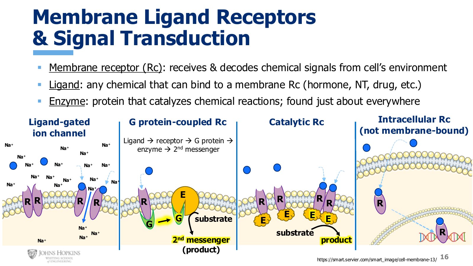
Slide 16. Four receptor arrangements. Only the first three are membrane-bound.
Ligand-gated ion channel. An ion channel, meaning a tunnel through the membrane, that stays closed most of the time and opens only when its specific ligand binds. Once open, ions pass through, here sodium. The ligand delivered the message to open the channel, because ion movement across the membrane is an important signal for how the cell should behave. This is how neurons and skeletal muscle cells work. Modules 2 and 3.
G protein-coupled receptor. The ligand binds the receptor, which activates an attached G protein. The G protein physically moves next door and binds a membrane-bound enzyme, changing that enzyme’s activity level. Since enzymes catalyze reactions, this changes the rate of substrate-to-product conversion. Increase the enzyme’s activity and you increase the product, a second messenger that travels through the cell telling it to change its behavior. Module 4.
Catalytic receptor. Nearly identical to a G protein-coupled receptor, except the enzyme is associated with the receptor directly. No G protein middleman needed.
Intracellular receptor. Not part of the membrane at all, but still a receptor where ligands bind. It is used only by hydrophobic solutes that cross the membrane easily on their own. The solute binds the intracellular receptor, which then crosses into the nucleus and affects how genes are expressed.
Nonpolar, polar, ionic
Transport proteins exist to help hydrophilic solutes cross, since water and anything that interacts easily with water cannot penetrate the hydrophobic core. So the question becomes how to tell which solutes are hydrophilic. It is easier to define hydrophilic by first defining its opposite.
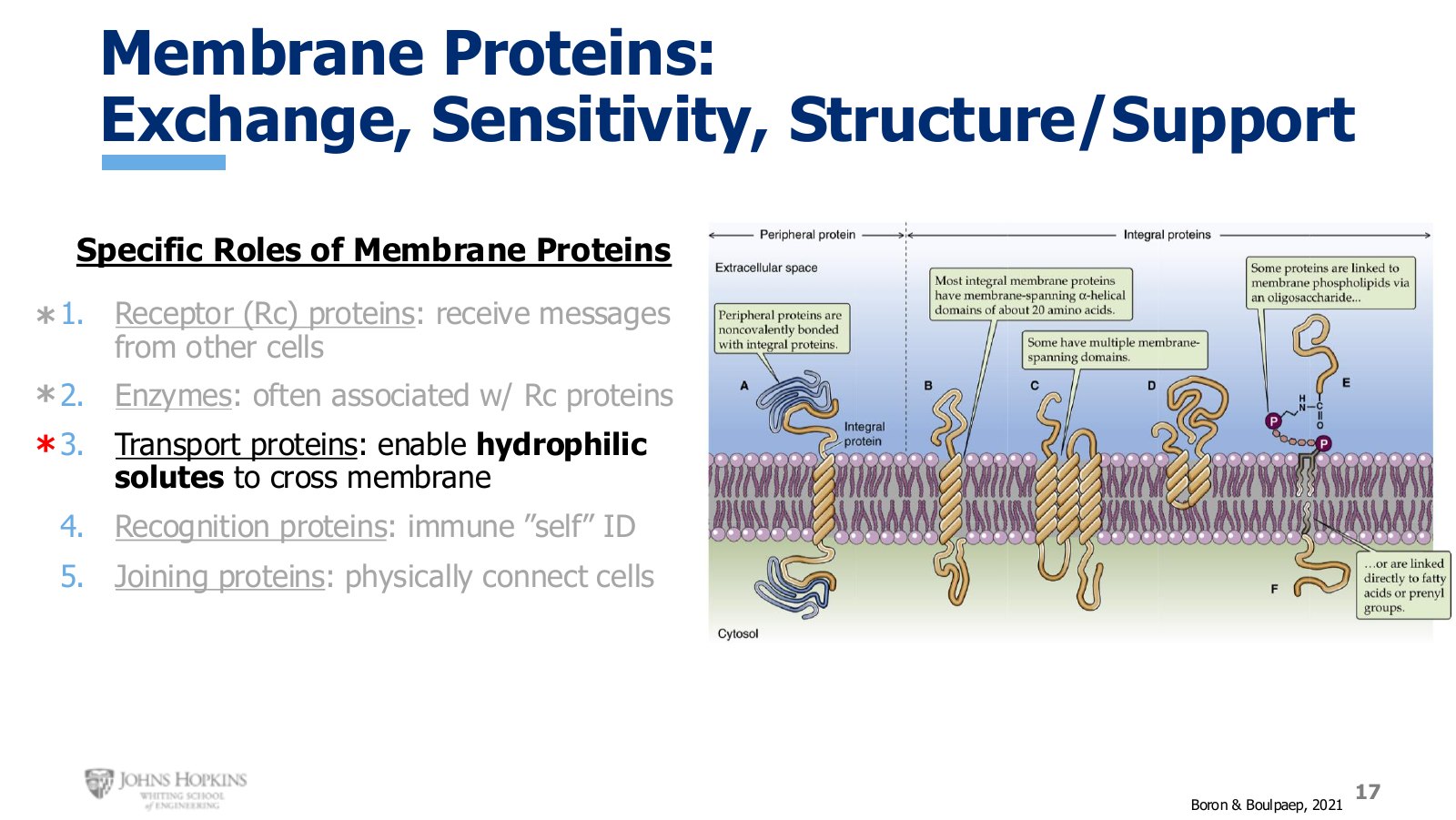
Slide 17. The five membrane protein roles again, now pointing at transport proteins and the solutes that need them.
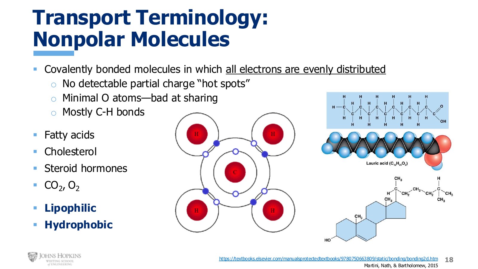
Slide 18. Nonpolar molecules. Lots of carbon-hydrogen bonds, very few oxygens.
A nonpolar molecule is covalently bonded, meaning held together by shared electrons, with all electrons fairly evenly distributed. Atoms share electrons so that each ends up with the right number to be more stable and less reactive, and that sharing binds them strongly.
Sometimes atoms share unequally, with one hugging the electrons more often. Oxygen is a big offender. But when sharing is even, as in methane, the negative charges carried by the electrons spread across the whole molecule and there are no detectable negative hotspots anywhere. No regional charge differences, no polarity.
Carbon and hydrogen are very good at sharing with each other, so any molecule built mostly of carbon-hydrogen bonds will be fairly nonpolar, especially with few oxygens. A fatty acid has two oxygens tacked on the end. Cholesterol has one. Other prominent nonpolar molecules in the body are steroid hormones like estrogen and testosterone, carbon dioxide, and, oddly enough, molecular oxygen.
Nonpolar means lipophilic and hydrophobic. These solutes cross the membrane easily without help, because their nonpolar nature lets them pass through the nonpolar fatty acid core.
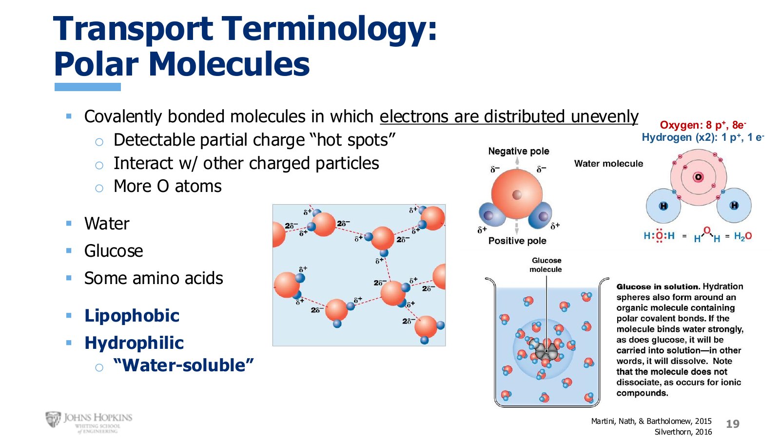
Slide 19. Water as the main example of a polar molecule, alongside glucose.
A polar molecule is also covalently bonded, but the atoms share badly. Electrons are unevenly distributed and one end holds them, and their negative charge, more often than the other. That produces detectable regional partial charges.
Water is the famous example. Oxygen already has eight electrons of its own, and once the two hydrogens share theirs to form the bonds, oxygen claims those shared electrons more strongly than hydrogen does. Electrons spend more time at the oxygen end, creating a negative partial charge there, a negative pole, while their absence at the hydrogen end creates a positive pole.
Those partial charges are what hold liquid water together. The slightly positive end of one water molecule is weakly attracted to the slightly negative end of another. These attractions are hydrogen bonds, and they produce cohesion and surface tension.
The same charges let water dissolve other polar molecules like glucose and some amino acids. The weak regional charges create handholds, giving water and solute a place to hold onto each other. Polar molecules are therefore lipophobic and hydrophilic, and they have a very difficult time crossing the membrane on their own.
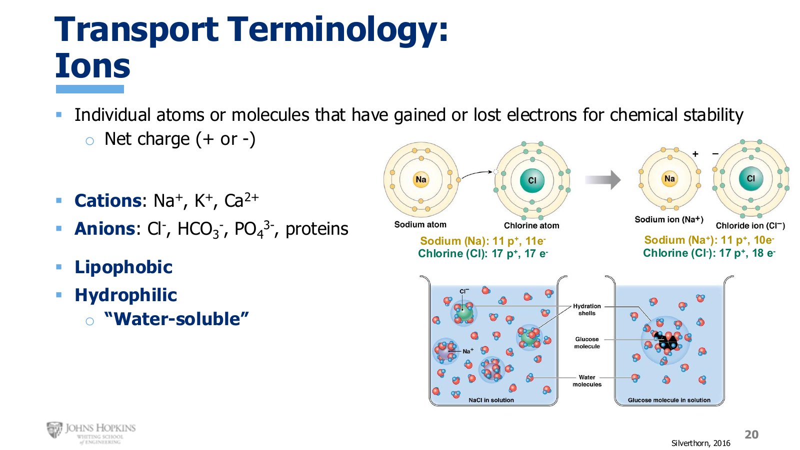
Slide 20. Sodium loses one electron, chlorine gains one. Both end up with a full outer shell and a net charge.
Ions behave differently again. Rather than sharing, an ion is typically only one or two electrons away from a full outer shell, so it simply loses or gains them.
Sodium and chlorine both have outer shells that hold up to eight electrons. Sodium has one electron there, chlorine has seven. Sodium’s solution is to lose that lone electron so the orbit disappears, leaving a full shell underneath. It keeps its 11 protons but drops to 10 electrons, giving a net charge of +1. Chlorine’s solution is to gain an electron someone else gave away, completing its outer shell of eight. It keeps 17 protons and now carries 18 electrons, a net charge of −1.
Unlike polar molecules, which have uneven internal charge distribution but no net charge, ions carry a net charge. The ones that matter this semester:
Cations (positive): Na+, K+, Ca2+
Anions (negative): Cl−, HCO3−, PO43−, proteins
Because ions are charged, they get along very well with water’s regional polarities. Water’s slightly positive hydrogen pole links up with negative ions like chloride, and its slightly negative oxygen pole links up with positive ions like sodium. Ions are lipophobic and hydrophilic, and they have an incredibly difficult time crossing the membrane on their own.