1E · Osmosis
Osmosis is a specialized type of diffusion: water molecules moving down a water concentration gradient toward concentration equilibrium.
We rarely think of water as having a concentration, so the usual framing is that water moves from an area with more water molecules and less solute, a less concentrated solution, toward an area with fewer water molecules and more solute, a more concentrated solution. It helps to picture water being pulled across the membrane toward the region of higher solute concentration.
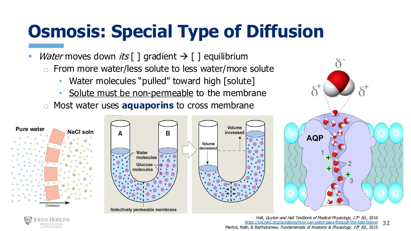
Slide 32. Osmosis into a NaCl solution, the U-tube demonstration, and aquaporins as the route water actually takes.
One condition governs everything: the solute must be unable to cross the membrane. If solutes cannot diffuse across to reach equilibrium themselves, water is forced to cross instead.
The U-tube shows it. A semi-permeable membrane sits in the middle. Small blue water molecules get through, large purple glucose molecules cannot. The left side holds dilute glucose, few purple molecules in many blue ones. The right side holds concentrated glucose, many purple in relatively few blue. Because the membrane is impermeable to glucose, water must move to equilibrate, so the level drops on the left and rises on the right.
Across a real cell membrane, water on its own has a hard time crossing the hydrophobic core. Cells rely on protein channels called aquaporins, literally water pores. A single aquaporin can pass a billion water molecules per second, which is why they are vital to osmosis in the human body.
Quantifying concentration
Because the concentrations of two adjacent solutions determine whether and in which direction osmosis occurs, we need units.
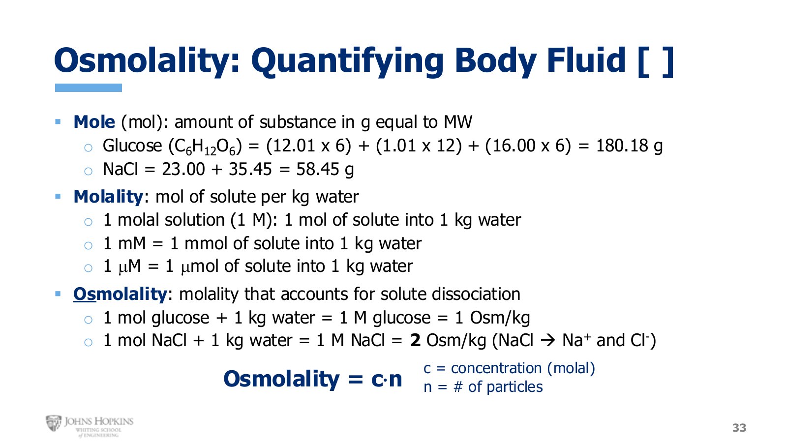
Slide 33. Mole, molality, osmolality. The glucose and NaCl worked examples and the osmolality = c · n relationship.
A mole of a substance is the amount of that substance in grams equal to its molecular weight. Two examples, using atomic masses from the periodic chart, which you do not need to memorize.
Molality expresses moles of a substance dissolved in one kilogram of water. A 1 molal solution, too concentrated for biological use, is one mole of solute in one kilogram of water. A 1 millimolal solution is one millimole in one kilogram, and so on down.
Osmolality is the term used in physiology textbooks. It takes into account how much of a solute dissociates into smaller particles once dissolved, so it quantifies how many solute particles are actually floating around.
Covalently bonded solutes like glucose do not dissociate, so no adjustment is needed. One mole of glucose in one kilogram of water gives an osmolality of 1 Osm/kg.
Ionically bonded molecules like magnesium sulfate, sodium bicarbonate, and sodium chloride break into their constituent ions on contact with water, because water breaks ionic bonds. One mole of NaCl in one kilogram of water is a 1 molal solution, but each NaCl molecule becomes two particles, a sodium ion and a chloride ion. Twice as many particles are floating in the water as were sitting on the scale, so multiply by two: an osmolality of 2 Osm/kg.
Osmotic gradients
Any difference in osmolality between two solutions is an osmotic gradient. Body fluid osmolality, particularly of the extracellular fluid, is regulated by negative feedback within a range of 280 to 310 mOsm/kg, targeting roughly 290.
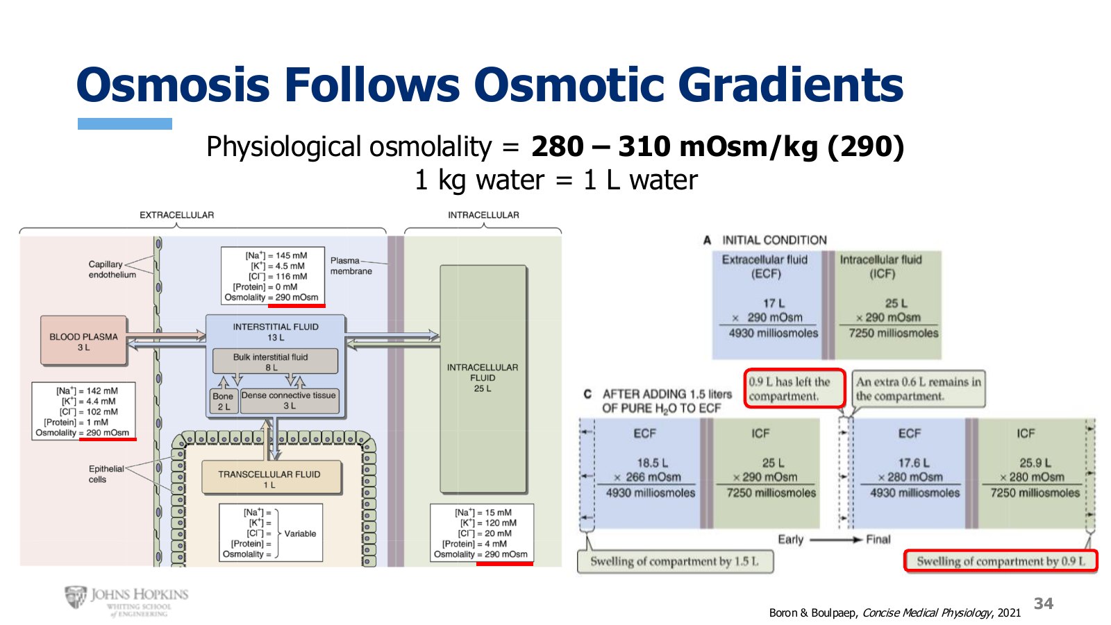
Slide 34. Plasma, interstitial, and intracellular compartments all sit at 290 mOsm in fluid homeostasis.
In fluid homeostasis, all compartments sit at 290 mOsm even though their solute makeup differs. Note that the “per kilogram” part of the unit is usually implied rather than written.
A worked perturbation, taking 1 kg of water as 1 L. At baseline the extracellular fluid holds 17 L at 290 mOsm/kg, so total solute is 17 × 290 = 4,930 mOsm. The intracellular fluid holds 25 L at 290.
Now infuse 1.5 L of pure water intravenously, which both expands and dilutes the extracellular fluid. Volume rises to 18.5 L, and concentration falls to 4,930 ÷ 18.5 = 266 mOsm/kg. An osmotic gradient now exists: the extracellular fluid is less concentrated than the intracellular fluid.
Water moves by osmosis from the extracellular into the intracellular compartment, a shift of 0.9 L, until both compartments sit at 280 mOsm/kg.
1.5 L pure water infusion, compartment by compartment
Stage | Extracellular volume | Extracellular osmolality | Intracellular volume | Intracellular osmolality |
|---|---|---|---|---|
Baseline | 17.0 L | 290 | 25.0 L | 290 |
Immediately after infusion | 18.5 L | 266 | 25.0 L | 290 |
After 0.9 L osmotic shift | 17.6 L | 280 | 25.9 L | 280 |
Osmotic pressure
Water movement is subject to more than osmosis. Hydrostatic pressure gradients also drive water in the body, sometimes in the same direction as osmotic gradients and sometimes against them. To compare their strengths directly, osmolality in mOsm/kg has to be converted into millimeters of mercury, a pressure unit.
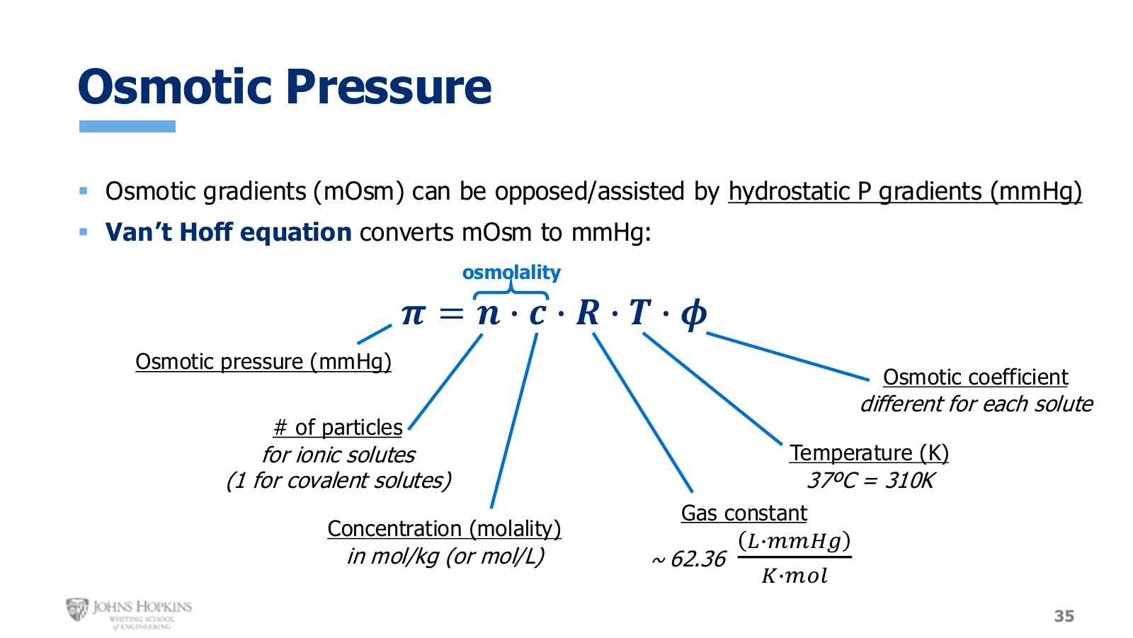
Slide 35. The Van’t Hoff equation, annotated term by term.
π is osmotic pressure, in mmHg.
n is the number of particles an ionic solute dissociates into. Use 1 for covalent solutes like glucose.
C is concentration expressed as molality, in mol/kg.
R is the gas constant, about 62.36 L·mmHg / K·mol. This is what introduces mmHg into the equation.
T is temperature in kelvin. Body temperature of 37°C is 310 K.
φ is the osmotic coefficient, different for every solute. You would be given this value.
The first two terms are just the osmolality expression from above. The last three are what convert it to pressure.
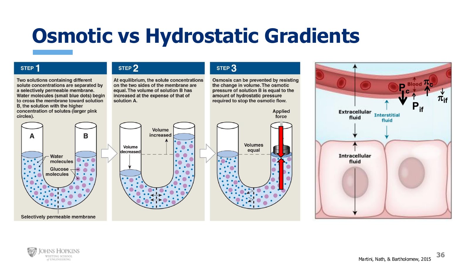
Slide 36. Osmotic and hydrostatic pressures in direct opposition, first in the U-tube and then across a capillary wall.
In the U-tube, a plunger pushes down on the right side, mimicking the hydrostatic pressure a column of water would generate. The downward hydrostatic pressure opposes the upward osmotic movement of water, and when the two are equal there is no net water movement.
The physiological version is water moving in and out of capillary blood vessels, where the capillary wall plays the role of the semi-permeable membrane. Four pressures act on the capillary at once, developed fully in Module 8.
The four pressures acting on a capillary
Pressure | Strength | Effect on water |
|---|---|---|
Capillary hydrostatic (Pc) | Strong | Pushes water out of the capillary |
Interstitial hydrostatic (Pif) | Weak | Pushes water into the capillary |
Capillary osmotic (πc) | Fairly strong | Pulls water into the capillary |
Interstitial osmotic (πif) | Weak | Pulls water out of the capillary |
Capillary osmotic pressure comes from non-permeable proteins in the plasma. Interstitial osmotic pressure comes from the very few proteins found in the interstitium. Two hydrostatic pressures push in opposite directions, layered on two osmotic pressures pulling in opposite directions. Because these forces change along the length of the capillary, the net effect on water movement changes from the beginning of the capillary to the end.
Three applications
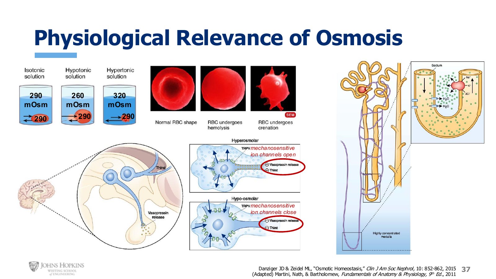
Slide 37. Red cells in three baths, a neuron opening and closing mechanically gated ion channels, and water recovery at the nephron.
Red blood cell shape. Net water movement across a membrane can radically change a cell’s shape.
A cell at 290 mOsm placed in a 290 mOsm bath shows no net water movement. Osmotic equilibrium. This is what the body aims for constantly.
In a less concentrated bath at 260 mOsm, water moves into the cell. Enough water and it bursts like a balloon, a process called hemolysis.
In a more concentrated bath at 320 mOsm, water moves out and the cell shrivels like a raisin, a process called crenation.
Structure dictates function, so when the physical structure of a red cell changes this way, the cell also stops working the way it needs to.
Neurons that sense dehydration. Neurons in the brain sense dehydration and respond by increasing body water: they prevent water loss at the kidneys through release of the hormone vasopressin, and they stimulate thirst to bring new water in. They detect dehydration by exploiting their own structural deformation.
When we are dehydrated, the fluid surrounding these cells is too concentrated, water leaves the neuron, and it shrivels. That shriveling physically opens mechanically gated ion channels, letting the neuron send electrical messages that stimulate vasopressin release and thirst. When the body is over-hydrated and the surrounding fluid is too dilute, water enters, the cell swells, the channels close, and the messages stop.
Water recovery at the nephron. The nephron is the microscopic functional unit of the kidney, and each kidney holds around one million of them. Water moves from the nephron back into interstitial fluid along an osmotic gradient set up next door by the NKCC cotransporter. As that cotransporter fills the interstitial space with extra ions, it concentrates the fluid, and the resulting osmotic gradient draws water out of the nephron and back into the body.