6A · General Electrophysiology Concepts: Review
This lecture briefly reviews the general electrophysiology concepts from earlier in the course so they are fresh, and adds heart-specific information along the way, tailoring the review to the cardiac emphasis of this module.
Nernst potentials
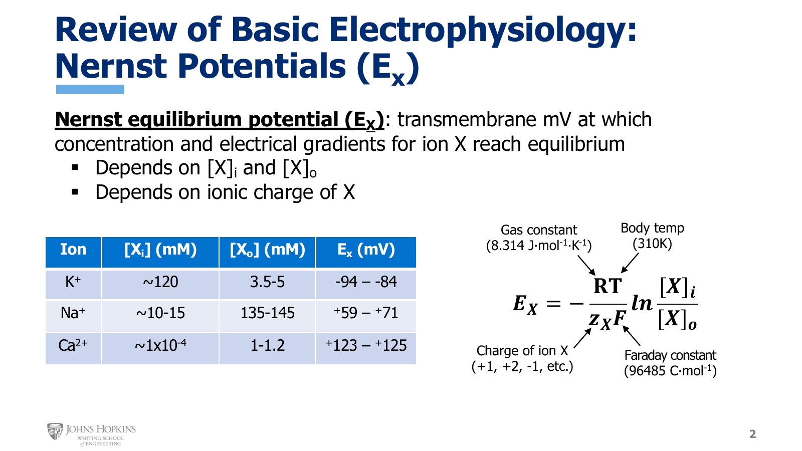
Slide 2. The Nernst equation with each constant labeled, and the intracellular and extracellular concentrations and Nernst potential ranges for K+, Na+, and Ca2+.
The Nernst potential is the membrane potential at which a given ion’s concentration gradient and electrical gradient have come to an equilibrium, and thus exert equal and opposite influences on the ion. At that millivoltage there is no net movement of that ion across the membrane. The equilibrium depends on the intracellular and extracellular concentrations of the ion and on the charge of the ion, all of which appear in the equation.
Multiply R, the gas constant (8.314 J·mol−1·K−1), by T, body temperature in Kelvin (310 K). Divide by the product of zX, the charge of the ion, and F, the Faraday constant (96485 C·mol−1). Make that value negative. Then multiply the whole term by the natural log of the ion’s intracellular concentration divided by its extracellular concentration.
The three ions of the cardiac action potential
Ion | [X]i (mM) | [X]o (mM) | EX (mV) |
|---|---|---|---|
K+ | ~120 | 3.5–5 | −94 to −84 |
Na+ | ~10–15 | 135–145 | +59 to +71 |
Ca2+ | ~1×10⁻⁴ | 1–1.2 | +123 to +125 |
Resting membrane potential
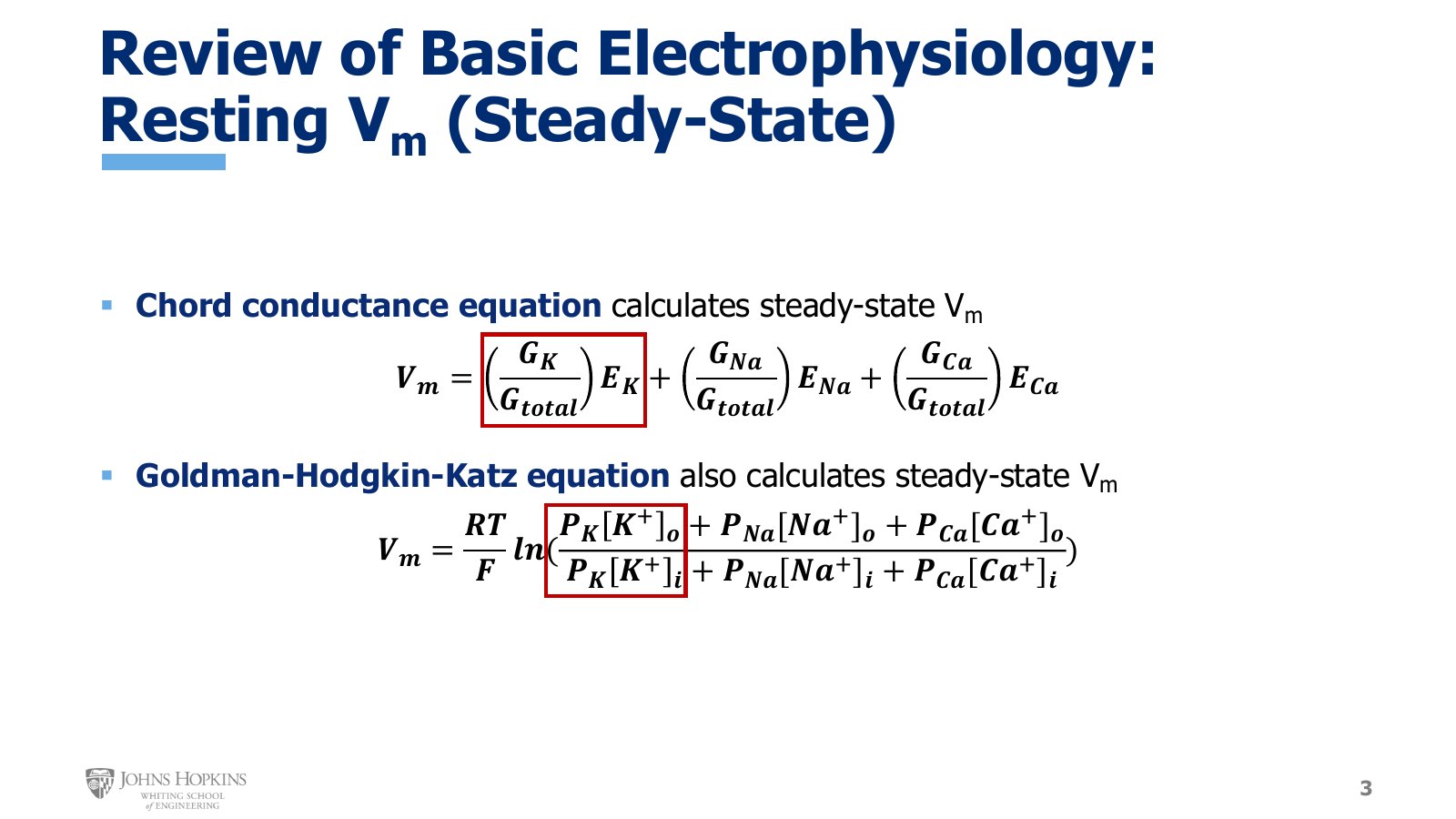
Slide 3. The chord conductance and Goldman-Hodgkin-Katz equations, with the potassium term boxed in each.
The chord conductance equation and the Goldman-Hodgkin-Katz (GHK) equation express the same idea. The resting membrane potential of a cell is determined by the Nernst potentials of all relevant ions, weighted by how much of each ion is able to cross the membrane under resting conditions.
In the chord conductance equation the Nernst potentials appear overtly as terms. Each is weighted by that ion’s conductance at rest divided by all ionic conductance at rest. Potassium ions are really the only ions that participate in any ion movement across the membrane at rest, so potassium conductance predominates and potassium exerts disproportional influence over resting potential.
In the GHK equation you need to recognize the Nernst equation inside it: RT divided by F, multiplied by the natural log of extracellular and intracellular ion concentrations, gives the Nernst potentials for each of the three ions. Each ionic term is multiplied by its permeability at rest. Potassium is the only ion with significant permeability at rest, so again the potassium term exerts disproportional influence over the final answer.
What Vm against EX tells you
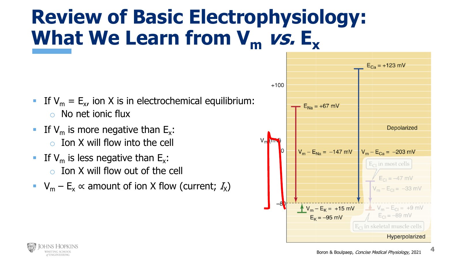
Slide 4. E-Na at +67 mV, E-Ca at +123 mV, and E-K at −95 mV against a resting potential of −80 mV, with a cardiac action potential sketched in red beside the scale.
Nernst potentials predict which direction an ion will move once you know the membrane potential. This familiar graph from the excitable membranes module now focuses on just the three ions of cardiac action potentials: sodium, potassium, and calcium. Four principles are the most practical.
If membrane potential equals the Nernst potential for an ion, there is no net movement of that ion across the membrane.
If membrane potential is more negative than the Nernst potential, sitting below it on the graph, the ion will flow into the cell. Cardiac action potentials range from as low as −90 mV up to around +30 mV. Sodium and calcium, with Nernst potentials up around +67 and +123 mV, always sit higher than any of those membrane potentials. Sodium and calcium will always want to flow into the cell.
If membrane potential is less negative than the Nernst potential, sitting above it on the graph, the ion will flow out of the cell. Potassium’s Nernst potential of −95 mV sits below the −80 mV resting potential on the graph, and below even the most negative −90 mV resting potential you might encounter. Potassium will always want to flow out of the cell.
The greater the difference between membrane and Nernst potentials, the stronger the ionic flow. Vm − EX is proportional to the current of ion X, IX.
Ionic currents in the heart
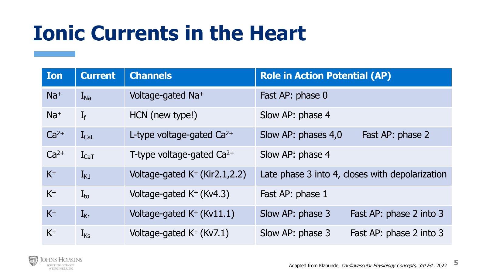
Slide 5. The eight cardiac currents with their ion, channel, and role in the slow and fast action potentials. A study table for the whole module.
Neural action potentials used only two kinds of ion channels, voltage-gated sodium and voltage-gated potassium, so there were really only two currents. The heart has many more types of ion channels. Each has its own unique characteristics, so the current through each has its own name and unique characteristics as well.
Sodium. INa is the standard sodium current through familiar voltage-gated sodium channels. If, the funny current, flows through a new type of channel, the HCN channel, which stands for hyperpolarizing cyclic nucleotide-gated.
Calcium. ICaL enters through L-type voltage-gated calcium channels, the calcium channels from the cardiac muscle lectures a couple of modules ago. ICaT enters through T-type voltage-gated calcium channels.
Potassium. Several currents, each through a different type of voltage-gated potassium channel. IK1 is also called the inward rectifying current. Ito stands for transient outward current. IKr and IKs stand for rapid and slow.
Cardiac ionic currents (slide 5)
Ion | Current | Channel | Role in the action potential |
|---|---|---|---|
Na+ | INa | Voltage-gated Na+ | Fast: phase 0 |
Na+ | If | HCN (new type) | Slow: phase 4 |
Ca2+ | ICaL | L-type voltage-gated Ca2+ | Slow: phases 4, 0. Fast: phase 2 |
Ca2+ | ICaT | T-type voltage-gated Ca2+ | Slow: phase 4 |
K+ | IK1 | Voltage-gated K+ (Kir2.1, 2.2) | Late phase 3 into 4, closes with depolarization |
K+ | Ito | Voltage-gated K+ (Kv4.3) | Fast: phase 1 |
K+ | IKr | Voltage-gated K+ (Kv11.1) | Slow: phase 3. Fast: phase 2 into 3 |
K+ | IKs | Voltage-gated K+ (Kv7.1) | Slow: phase 3. Fast: phase 2 into 3 |