6C · Slow-Response Cardiac Action Potential
This lecture covers the slow response waveform and its phases, its ionic currents and channels, the tissues that show it, and its significance in overall cardiac function.
The waveform
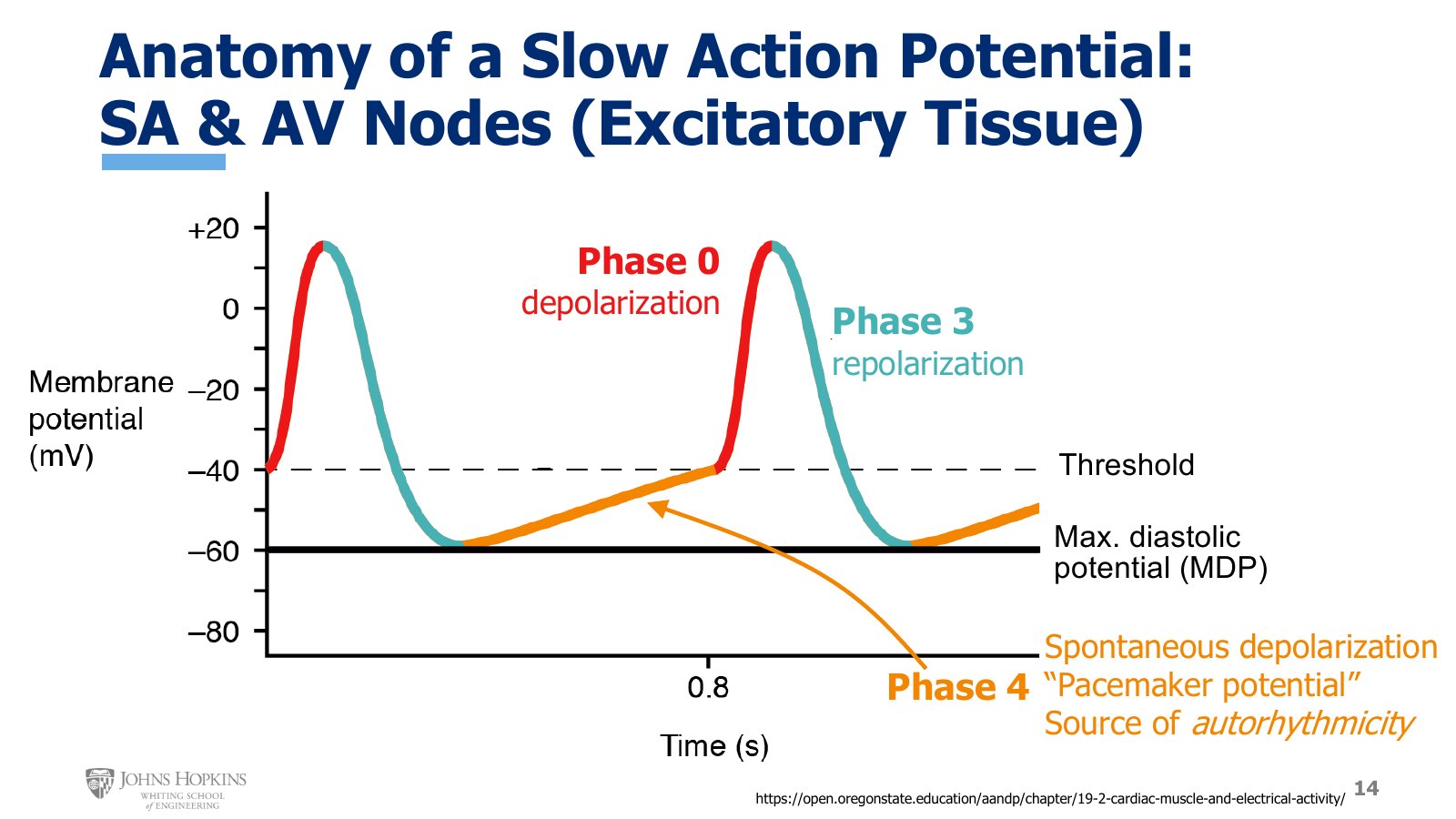
Slide 14. The slow action potential: phase 0 depolarization in red, phase 3 repolarization in teal, and phase 4 creeping from the maximum diastolic potential near −60 mV up to threshold near −40 mV.
The slow response action potential is what makes excitatory tissues excitatory. It appears in both nodes of the heart: the SA node as primary pacemaker and the AV node as secondary pacemaker.
It has only three phases. Phases 1 and 2 do not appear in this waveform, so they are skipped.
Phase 4, a resting phase that is not truly resting, because it is slowly creeping up.
Phase 0, depolarization.
Phase 3, repolarization.
The creeping phase 4 gives this action potential its pacemaker function. It slowly, gradually depolarizes from its most negative point, around −60 mV, called the maximum diastolic potential (MDP), up to threshold around −40 mV, on its own. The slow depolarization to threshold is spontaneous: the cell does it by itself, without any external influence.
Because it is so critical to the excitatory function of the SA and AV nodes, phase 4 has a few names. Some call it spontaneous depolarization, which is very descriptive. Almost all textbooks call it the pacemaker potential. Whatever the name, it is the source of the heart’s autorhythmicity, its ability to depolarize itself and so set its own rhythm of beating.
Phase 4 currents
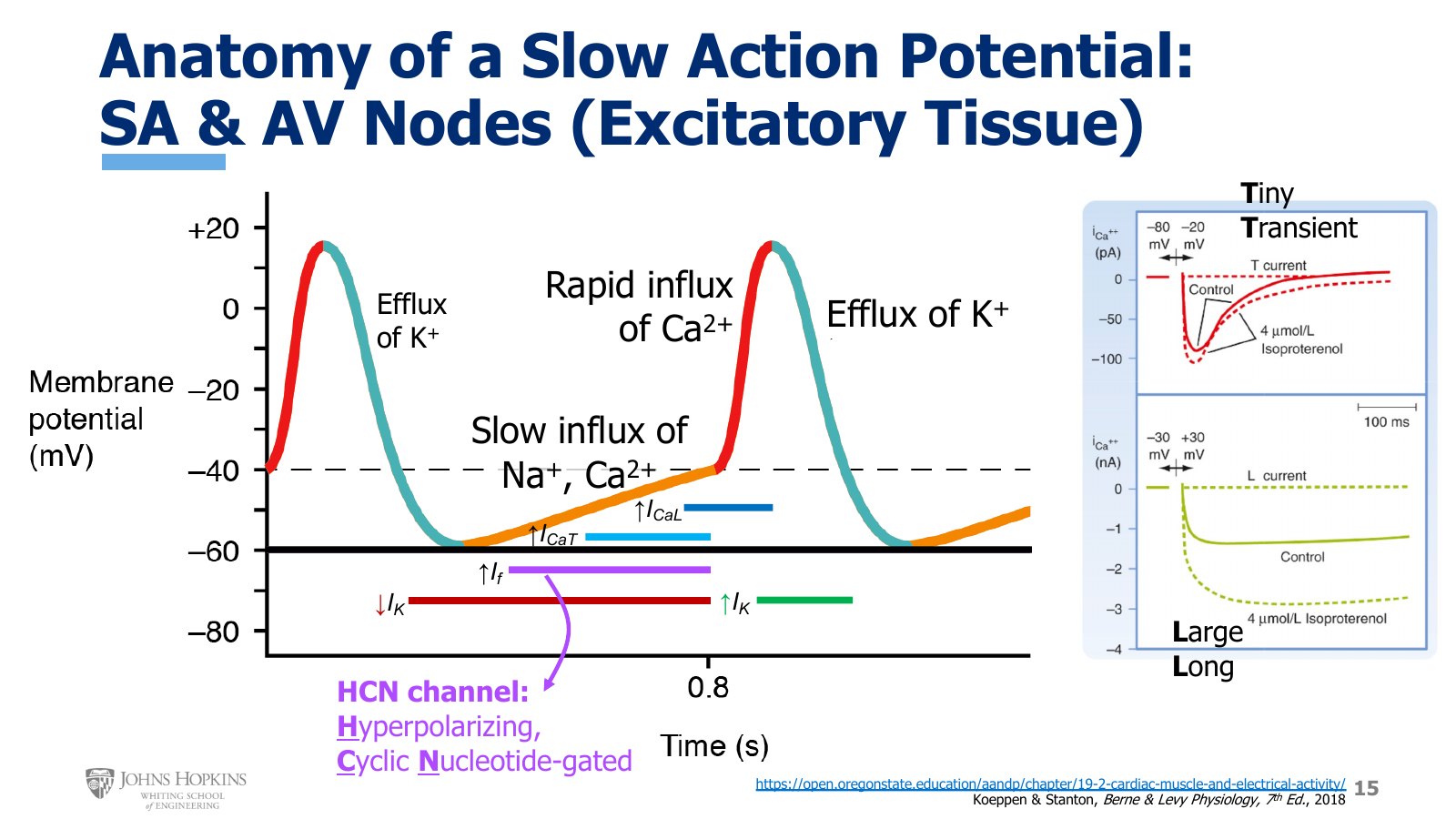
Slide 15. The currents of each phase drawn as bars under the waveform: falling I-K, then I-f, I-CaT, and I-CaL in phase 4, rising I-K in phase 3. At right, T-type current (tiny, transient, in pA) against L-type current (large, long, in nA).
Start with phase 4, because it is both the most involved and the most unusual. Four different ionic currents contribute to its slow upward creep.
Decreasing potassium efflux. Phase 3 repolarization is driven by potassium efflux. Near the end of phase 3 and well into phase 4, potassium efflux slows down continuously. Fewer positive charges are lost, and at a much slower rate, which helps turn the corner from repolarization into depolarization.
Funny current, If. Reaching the maximum diastolic potential serves as the stimulus for the funny current, mostly sodium influx through HCN channels (hyperpolarizing cyclic nucleotide-gated). Most voltage-gated channels open in response to depolarization. HCN channels open in response to hyperpolarization, so the most negative potential is the stimulus that opens them. That hyperpolarizing stimulus struck the scientists who discovered the current as strange, weird, funny, which is where the funny current gets its name.
T-type calcium current, ICaT. Once the gentle inward sodium flow of the funny current has brought membrane potential up a little, T-type calcium channels open. T-type calcium current flows in and further depolarizes the membrane.
L-type calcium current, ICaL. That in turn opens L-type calcium channels. L-type calcium current flows in, the final step in bringing membrane potential all the way up to threshold.
Phase 4 is involved, with several currents at work, but it is carefully orchestrated to bring the cell to threshold in a calculated, predictable, regulated manner.
Phases 0 and 3
At threshold most of the phase 4 currents disappear, but not the L-type calcium current. Reaching threshold opens more voltage-gated L-type calcium channels, so this current becomes the major driving force behind phase 0 depolarization. This is unique. In most other action potentials, sodium influx drives depolarization. Here it is calcium-driven.
Once the calcium channels start to close, voltage-gated potassium channels open and potassium flows out of the cell, repolarizing it during phase 3. Then it all starts again.
T-type against L-type calcium channels
Most of the time in excitable membranes the calcium channel in question is L-type. Phase 4 of the slow action potential is really the only time T-type channels get involved, and they are different. On the graph, T-type calcium influx is on top and L-type on the bottom.
T-type channels do not allow much calcium through (the units are picoamps), and the influx does not last long. T stands for tiny conductance, transient opening.
L-type channels produce a much larger calcium influx, measured in nanoamps rather than picoamps, and the inward current lasts much longer. L stands for large conductance, long-lasting opening.
The SA node
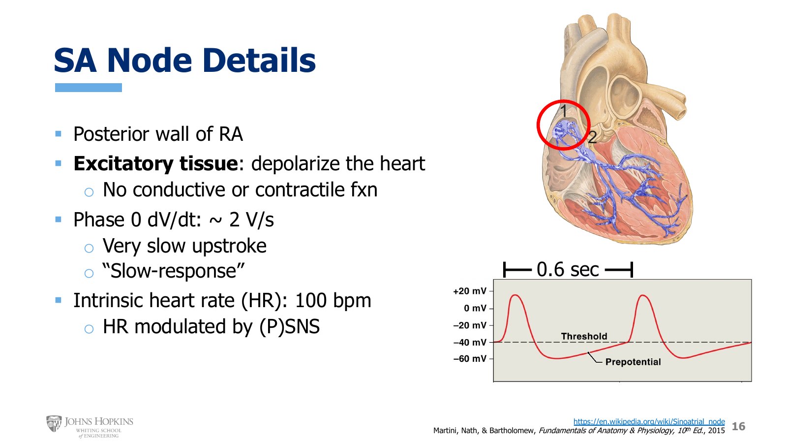
Slide 16. The SA node, circled, high on the posterior wall of the right atrium. Its slow waveform repeats every 0.6 s, with the gently rising phase 4 labeled prepotential.
Location. The posterior wall of the right atrium, on the back, up near the top of the chamber.
Role. Excitatory tissue, the main pacemaker of the heart. Its job is to send regularly timed waves of depolarization to the rest of the heart. It has no conductive or contractile function.
Upstroke. Phase 0 is quite gradual. Its dV/dt is approximately 2 V/s, by far the slowest rate of depolarization anywhere in the heart, hence the term slow response action potential.
Rate. The SA node depolarizes spontaneously about every six tenths of a second, around 100 times per minute.
That gives an intrinsic heart rate of about 100 beats per minute. The intrinsic heart rate is simply how often the heart beats on its own in the absence of any neural input. The frequency of depolarization can be modulated by input from the sympathetic and parasympathetic nervous systems.
Chronotropy: modulating heart rate
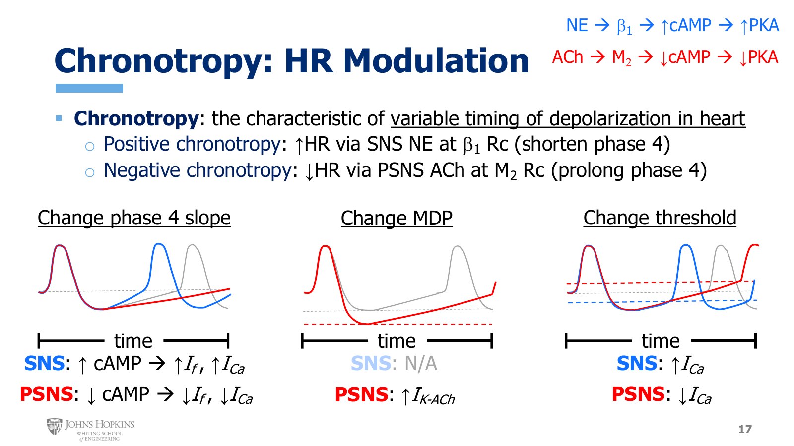
Slide 17. Three ways to change phase 4 duration: its slope, the maximum diastolic potential, and threshold. Blue traces are sympathetic, red parasympathetic, gray baseline.
Chronotropy is a term unique to the heart. Chrono- means time, and chronotropy describes the inherently variable timing of depolarization in the heart: the frequency of depolarization in the SA node, and so heart rate, is variable. It is the first of four tropic terms in the cardiovascular system, all of which reflect inherently variable characteristics of the heart.
Positive chronotropy is an increase in heart rate. Sympathetic nerves release norepinephrine onto beta-1 receptors on the SA node. The overall effect is to shorten phase 4: less time between depolarizations, less time between heartbeats, a faster heart rate.
Negative chronotropy is a decrease in heart rate. Parasympathetic nerves release acetylcholine onto M2 receptors on the SA node. The overall effect is to prolong phase 4: more time between depolarizations, more time between heartbeats, a slower heart rate.
Phase 4 duration changes in three ways: by changing the slope of phase 4, by changing the maximum diastolic potential (the most negative millivoltage before phase 4 starts), and by changing where threshold sits.
1. Slope of phase 4. Recall from cardiac muscle that sympathetic norepinephrine binds beta-1 receptors, increasing cyclic AMP production, which increases protein kinase A activity, which led to intracellular events that increased force of contraction. Acetylcholine binding muscarinic receptors did the opposite. The SA node shows the same changes in cyclic AMP and PKA, but the intracellular effects differ, because the SA node is not contractile tissue.
Sympathetic. Norepinephrine increases cyclic AMP, which directly increases sodium influx through the funny current, because HCN channels are stimulated by cyclic nucleotides like cyclic AMP. More cyclic AMP, more open HCN channels, more funny current, faster depolarization, a steeper slope. Threshold arrives sooner and phase 4 shortens. More cyclic AMP also means more PKA, which phosphorylates calcium channels and opens them, giving more calcium current, again faster depolarization and a steeper slope.
Parasympathetic. Acetylcholine decreases cyclic AMP and PKA, decreasing both the funny current and the calcium current. Depolarization during phase 4 slows, the slope flattens, and reaching threshold takes longer.
2. Maximum diastolic potential. Only the parasympathetic side changes this; there does not seem to be a good sympathetic correlate. Specific potassium channels open only when acetylcholine is present. The potassium flow through them is the IK-ACh current, and it is especially active during phase 3. More potassium efflux than normal during phase 3 makes the most negative membrane potential even more negative. When the waveform starts creeping back up in phase 4, it has much farther to go before hitting threshold, so it takes longer to get there: a longer phase 4.
3. Threshold. Through mechanisms still somewhat debated, changes in the inward calcium currents can change where threshold sits. The same calcium current changes from the slope effect are at work here too.
A sympathetic increase in calcium influx shifts threshold downward, so it sits very close to the maximum diastolic potential. The membrane does not have to travel far before hitting threshold, shortening phase 4.
A parasympathetic decrease in calcium influx shifts threshold upward, so it sits much farther from the maximum diastolic potential than before. The membrane has farther to travel, which takes longer and prolongs phase 4.
The AV node
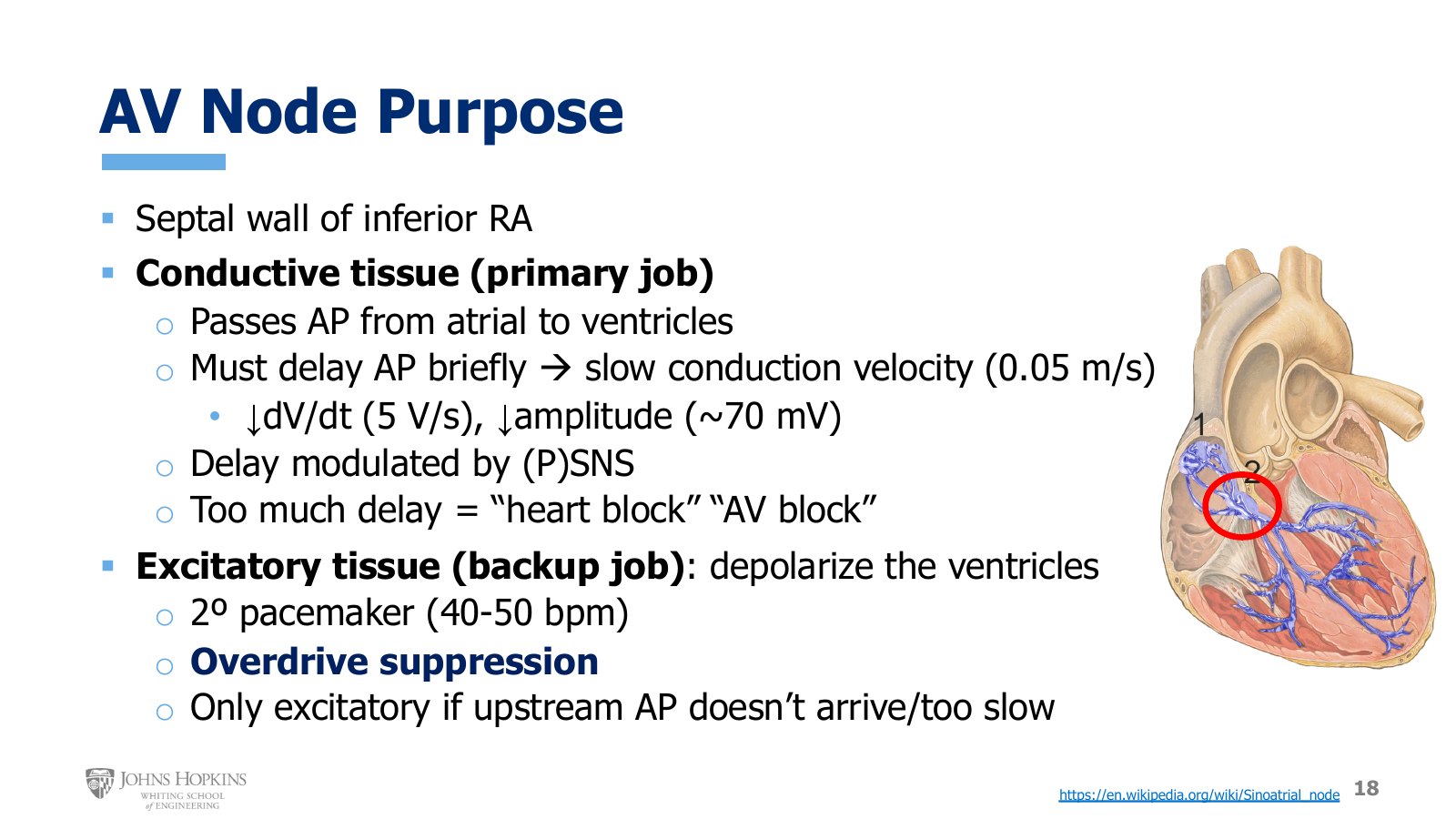
Slide 18. The AV node, circled, low on the septal wall of the right atrium, with its primary conductive job and backup excitatory job.
The AV node sits at the bottom of the right atrium, over by the septal wall.
Primary job: conductive. It collects the action potential after it passes through the atria and sends it down to the bottom half of the heart. It is the lone point of electrical access into the ventricles. A critical part of its conductive function is to delay the action potential briefly, for about a tenth of a second, which lets the atria finish depolarizing before the signal goes down to the ventricles. Conduction through the atria is around 0.5 m/s. The AV node delays transit by dramatically slowing conduction to about 0.05 m/s, ten times slower than in the atria. It does this through a combination of a low phase 0 dV/dt, only about 5 V/s, and a fairly small overall amplitude, only about 70 mV from top to bottom.
The delay can be modulated by the sympathetic and parasympathetic nervous systems. If the delay becomes too dramatic or prolonged, it can contribute to a condition known as heart block or AV block, where the AV node fails to pass the action potential to the ventricles.
Backup job: excitatory. With a slow action potential and a sloping phase 4, the AV node can spontaneously depolarize, but much more slowly than the SA node: only about 40–50 times per minute, because it has fewer HCN channels and so a weaker funny current. Normally the much faster depolarization arriving from the SA node suppresses the AV node’s pacemaker activity through a phenomenon called overdrive suppression, forcing the AV node to march to a much faster incoming beat. If upstream action potentials from the SA node do not arrive in a timely manner, due to SA node or atrial dysfunction, AV node pacemaker activity takes over as the primary pacemaker for stimulating the ventricles. It is not ideal and requires medical attention, but the AV node can step in when the SA node fails.
Dromotropy: modulating conduction velocity
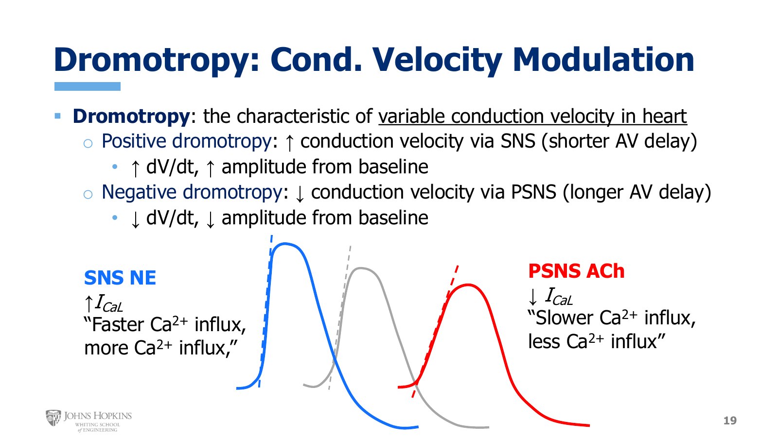
Slide 19. Against the gray baseline, sympathetic norepinephrine gives a steeper upstroke and taller peak (blue). Parasympathetic acetylcholine gives a gentler upstroke and lower peak (red).
Dromotropy is the second tropic term for the heart. It refers to the inherently variable conduction velocity through the heart, which neural input can manipulate and modulate. Dromo- is a Greek root referring to a race.
Positive dromotropy is an increase in conduction velocity, as the action potential races through the AV node more quickly. The shorter AV delay comes from a sympathetically induced increase in both the phase 0 dV/dt and the overall amplitude of the waveform.
Negative dromotropy is a decrease in conduction velocity, giving a longer AV delay. It comes from a parasympathetically induced decrease in both the phase 0 dV/dt and the overall amplitude.
If the gray waveform is the baseline, sympathetic positive dromotropy shows a steeper dV/dt upslope in phase 0 and a taller peak millivoltage. Sympathetic stimulation opens more L-type calcium channels, just as it did in the SA node. Faster calcium influx makes dV/dt steeper, and more overall calcium influx gives a higher peak by the time those channels close.
Parasympathetic negative dromotropy shows a gentler dV/dt upslope in phase 0 and a depressed peak. Parasympathetic stimulation closes some calcium channels, just as it did in the SA node. Slower calcium influx gives lower dV/dt, and less overall calcium influx gives the lower peak amplitude.