7A · Heart Anatomy
This lecture covers the major chambers, valves, and vessels associated with the heart.
Chambers
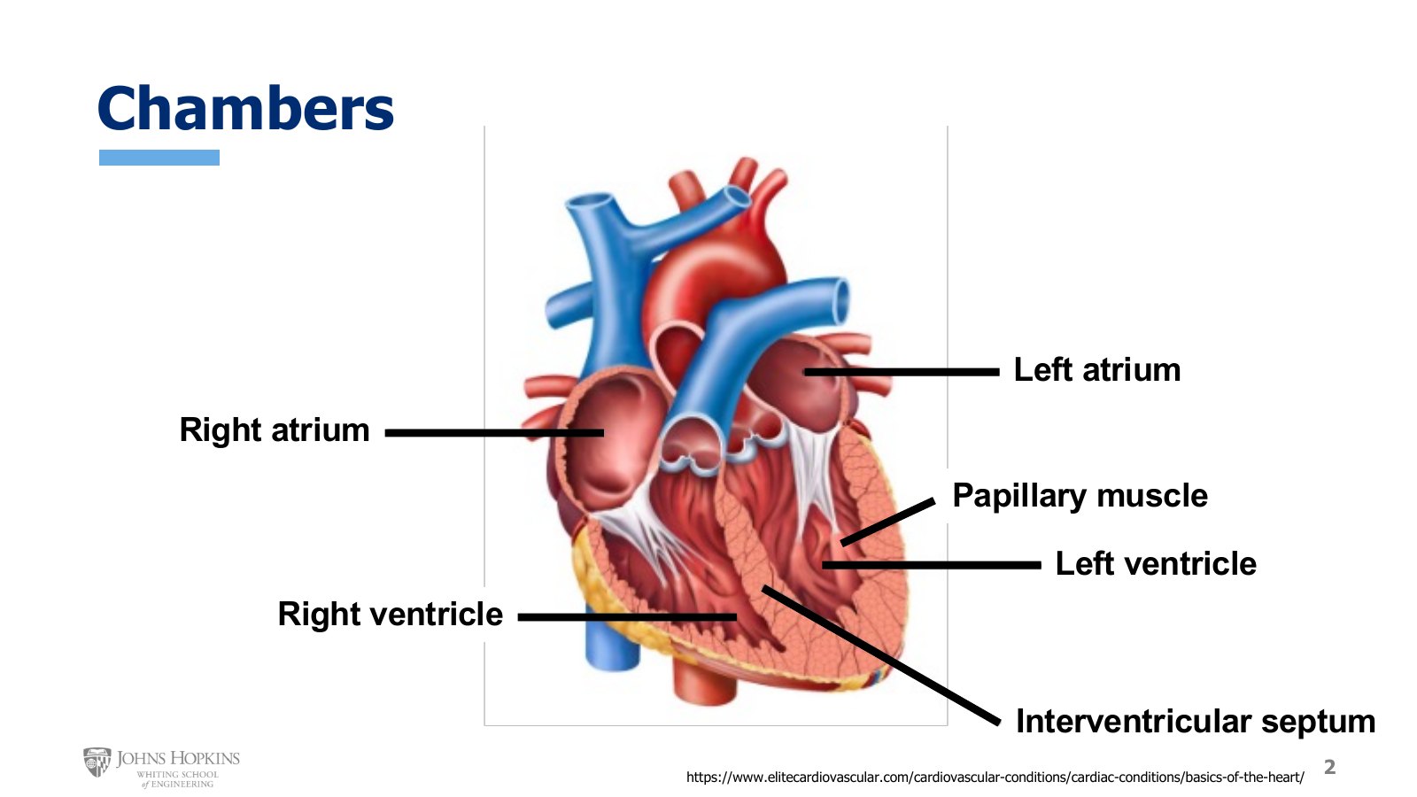
Slide 2. The four chambers, with the interventricular septum dividing the ventricles and papillary muscles reaching into the left ventricle.
The four chambers are review from the module on cardiac electrophysiology, and good to keep fresh in your mind. On the right side of the heart, the left side of the figure, the right atrium sits on top with the right ventricle below it. On the left side of the heart, the right side of the figure, the left atrium sits on top with the left ventricle below it.
Two more structures go with the chambers. The interventricular septum is the thick wall separating the two lower chambers. It keeps the low pressure deoxygenated blood of the right ventricle separated from the high pressure oxygenated blood of the left ventricle.
The unusual muscular outcroppings extending from the ventricular wall into the lumen of the chamber are the papillary muscles, named because they resemble papillae, or fingers. They provide structural stability to some of the heart valves.
Valves
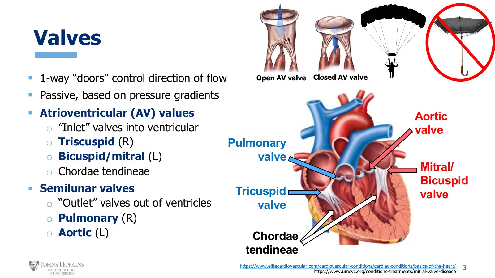
Slide 3. The four valves and the chordae tendineae. An open and a closed AV valve, a parachute held by its cords, and an umbrella blown inside out.
The valves are new for us. Much like there are four chambers, there are four valves, and they work as one-way doors that control the direction and timing of blood flow as blood moves from chamber to chamber and from chamber to vessel. The valves contain no muscle tissue at all. They are passive structures in that sense, opening and closing purely on the prevailing pressure gradients around them.
The atrioventricular, or AV, valves control the flow of blood from the atria into the ventricles, functioning as inlet valves into the ventricles.
Right side: the tricuspid valve, with three cusps, or flaps.
Left side: the mitral valve, or bicuspid valve. Bicuspid is the more anatomically descriptive name, since the valve has only two cusps. Mitral is the term you will likely hear more often, particularly with clinical conditions like mitral stenosis or mitral insufficiency.
The fibrous strings, or cords, connected to the AV valves are the chordae tendineae. Their job is to connect the edges of the AV valves to the papillary muscles.
As the inlet valves for the ventricles, the AV valves need to be open to let blood flow down into the ventricles, but they need to close and stay closed while the ventricles contract and generate pressure to pump blood out through a different valve. The problem is that the ventricle generates significant pressure when it contracts, which could make the AV valves prolapse, or blow out backwards like an umbrella turned inside out in a windstorm. That would damage the valves and create ongoing valve disease because of the structural instability.
The chordae tendineae prevent this by holding onto the edges of the valve. When the papillary muscle at the other end of the chordae contracts and pulls down, the chordae pull down on the valve edges and keep the valve from prolapsing. It is a bit like the cords on a parachute. Cords attached to the edges let the parachute inflate and catch the air, much like chordae attached to the valve edges let it catch the upward movement of pressurized blood without blowing out like an umbrella.
The other two valves are the semilunar valves, the outlet valves for the ventricles, which open when the ventricle contracts and generates pressure.
Right ventricle: the pulmonary valve.
Left ventricle: the aortic valve, which tends to hide behind the pulmonary valve.
The valves from above
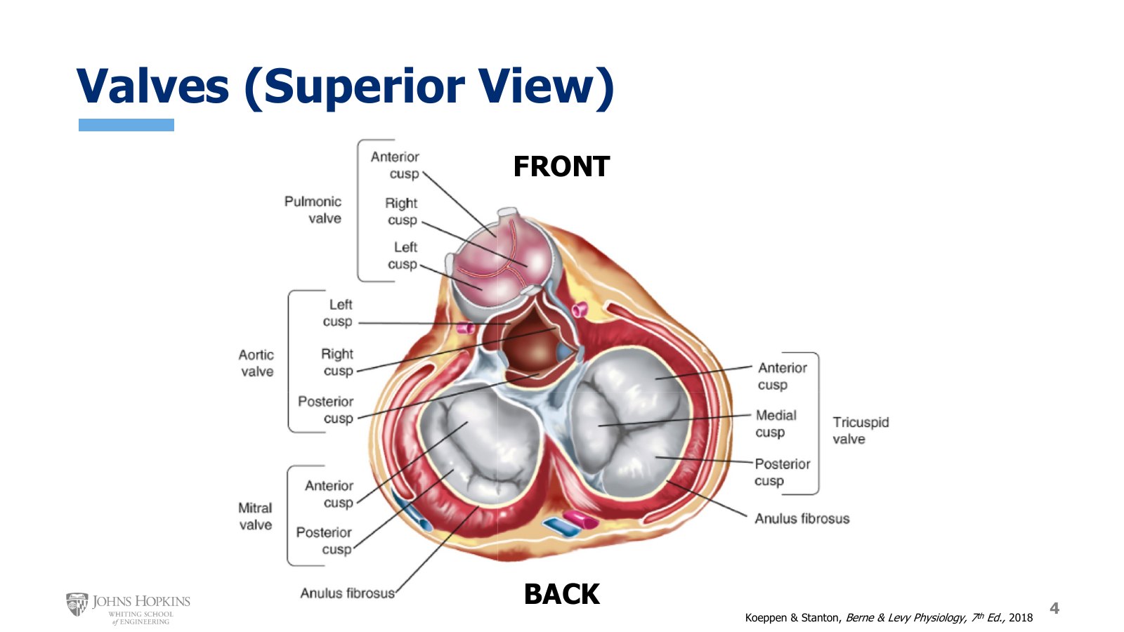
Slide 4. The atria removed and the ventricles viewed from the top, front of the heart at the top of the image. The slide labels the pulmonary valve as the pulmonic valve.
Here is a different perspective to help you assemble a three-dimensional picture of the heart in your head from multiple two-dimensional images along different planes. The atria have been removed from the top of the heart, and we are looking down on the top of the ventricles. The back of the heart is at the bottom of the image and the front is at the top.
The tricuspid valve on the right side of the image, with its three flaps.
The mitral, or bicuspid, valve on the left side of the image, with its two flaps.
The pulmonary valve in front, with three cusps: right, left, and anterior, or front.
The aortic valve hiding behind it, also with three cusps: left, right, and posterior, or back.
Valve disease: stenosis and regurgitation
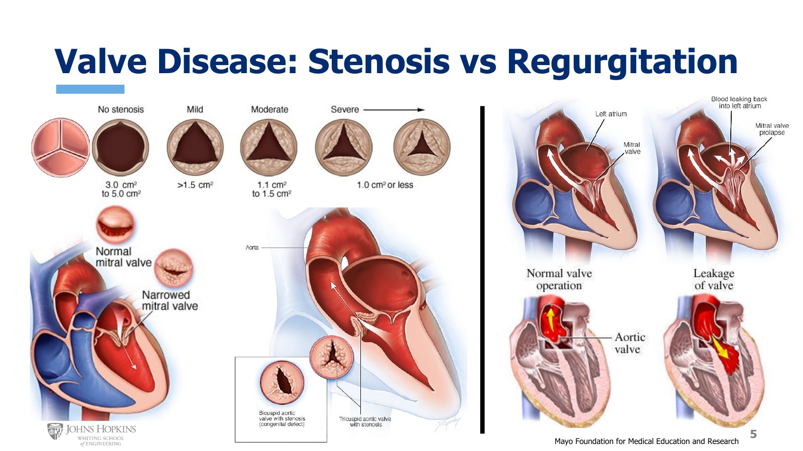
Slide 5. Left, aortic stenosis graded from none to severe, a narrowed mitral valve, and a bicuspid aortic valve. Right, mitral and aortic regurgitation against normal forward flow.
Valvular disease is incredibly common, and developing innovative solutions for diseased valves is a prominent element of cardiovascular engineering. It tends to fall into one of two categories: stenosis or regurgitation. We typically see valvular disease in the valves of the left heart, mitral and aortic. That side of the heart experiences higher pressures, so there is simply more wear and tear on those tissues.
Valve stenosis occurs when the valve cusps become calcified over time, making the valves harder to open and close and narrowing the valvular opening. The top of the slide shows a typical age-related progression of stenosis, or narrowing, of the aortic valve.
A healthy valve with no calcification looks smooth and opens fully, with an opening of 3 to 5 cm2.
With the high pressures and high blood velocities inherent to the left side of the heart, the aortic valve eventually develops some tissue damage and scar tissue, which leads to a little calcification, visible as little bumps.
Calcification makes it harder to open the valve fully, which leads to a slight narrowing.
Narrowing increases blood velocity more, further damaging the valve and leading to more scarring, calcification, and narrowing.
The cycle continues over time until the stenosis is so severe that the heart cannot pump enough blood out to the body anymore. The opening is less than 1 cm2.
At that point the typically elderly patient goes into symptomatic heart failure, which requires surgery to replace the valve.
Lower left is a stenotic mitral valve, severely limiting blood flow from the left atrium into the left ventricle. Beside it is a common variation of aortic stenosis, the bicuspid aortic valve. Two cusps were fused as a birth defect, creating abnormal flow patterns through the valve and resulting in calcification and stenosis at much younger ages.
On the right side of the slide is valvular regurgitation, or insufficiency, compared with normal forward flow. Regurgitation occurs when a damaged valve allows blood to flow backward, backwashing into the chamber from which it just came.
Mitral regurgitation occurs while the left ventricle is contracting and trying to eject blood through the aortic valve. Some blood flows backward through the mitral valve into the left atrium, because the chordae tendineae are not doing a good job of keeping the valve closed during contraction.
Aortic regurgitation occurs after the left ventricle has finished contracting and the aortic valve is supposed to be closed. Some of the blood just ejected through the valve flows backward into the left ventricle again.
Regurgitant valves mean that insufficient blood volume ends up moving in a net forward direction. The chambers that experience the backflow end up with pathologically higher volumes over time, which can lead to chronic complications like heart failure or arrhythmias.
Great vessels
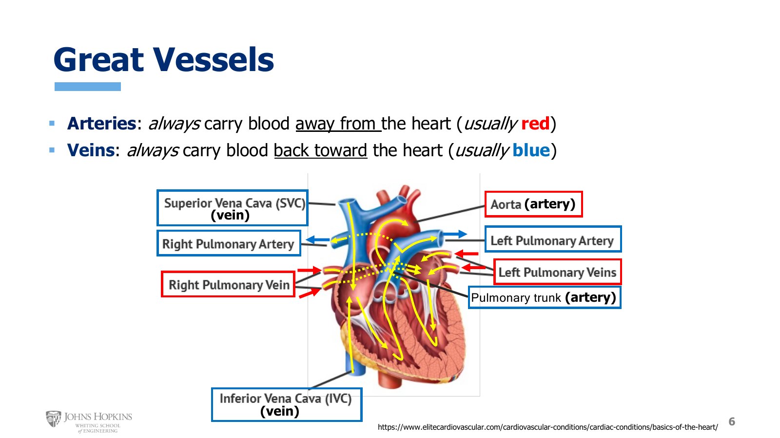
Slide 6. The great vessels, boxed red or blue by the blood they carry and labeled artery or vein by direction. Yellow arrows trace flow through the heart.
The great vessels are the blood vessels that lead directly into or out of the chambers of the heart. First we need the meaning of arteries and veins.
Arteries are usually red, carrying oxygenated blood, but there are exceptions, some of them on this figure. The definition that is true 100% of the time: arteries carry blood away from the heart. Likewise, veins are usually blue, carrying deoxygenated blood, with exceptions here too. The definition that is true 100% of the time: veins carry blood back toward the heart.
The superior vena cava is the vein bringing deoxygenated blood back from the head, neck, arms, shoulders, and upper chest, draining down into the right atrium from above.
The inferior vena cava is the vein bringing deoxygenated blood back from the lower chest, abdomen, pelvis, and legs, draining into the right atrium from below.
From the right atrium, blood flows through the tricuspid valve into the right ventricle, then out through the pulmonary valve into the pulmonary trunk. The pulmonary trunk is considered an artery because it carries blood out of and away from the heart.
The pulmonary trunk branches into the left and right pulmonary arteries, carrying deoxygenated blood to the left and right lungs.
After the blood travels to the lungs and becomes oxygenated, it returns through the left and right pulmonary veins and enters the left atrium.
From the left atrium it travels through the mitral valve into the left ventricle, then out through the aortic valve into the aorta, the main artery of the circulation, carrying blood from the heart to the tissues of the body.
Pressure gradients in the heart
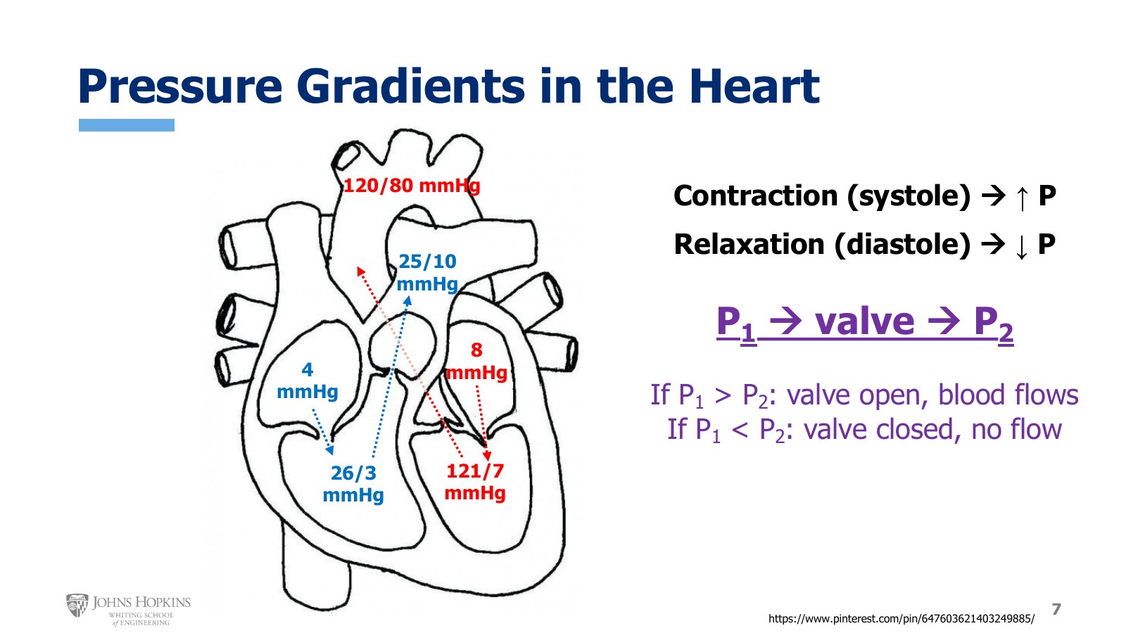
Slide 7. Typical pressures in each chamber and outflow vessel, with the rule for when a valve opens.
Consider blood flowing from region one, with pressure P1, through a valve to region two, with pressure P2. How P1 compares with P2 determines whether the valve is open or closed.
If P1 > P2, the valve opens and blood flows from region one to region two.
If P1 < P2, the valve is closed and no blood can flow.
The pressures in the chambers and great vessels are not constant. They can be quite variable, which is why valves spend some time open and the rest of the time closed. Pressures change because the heart contracts. Contraction in the heart is systole, and when contraction occurs, particularly in the ventricles, pressure increases. Relaxation in the heart is diastole, and when relaxation occurs, particularly in the ventricles, pressure decreases. Because vessels are attached to the ventricles, the vessels also experience pressure changes tied to the upstream ventricular systole and diastole.
These pressures vary, sometimes widely, from person to person, but they are a reasonable place to start for a general sense of heart and arterial pressures.
Typical pressures, systolic/diastolic where they swing
Location | Pressure (mmHg) |
|---|---|
Right atrium | ~4 |
Right ventricle | 26/3 |
Pulmonary trunk | 25/10 |
Left atrium | ~8 |
Left ventricle | 121/7 |
Aorta | 120/80 |
Atrial pressure does not fluctuate much between systole and diastole, so the atria get average values. Right atrial pressure sits around 4 mmHg, and left atrial pressure a smudge higher, around 8.
Ventricular pressures are a different story. The right ventricle gets as low as 3 mmHg when relaxed and as high as 26 when contracted. The pulmonary trunk sees a similar range: 10 mmHg when the right ventricle is in diastole and 25 when it is in systole.
The left side tells the same story, with the only difference being much higher systolic pressure, because the thicker, stronger wall of the left ventricle generates more pressure. Left ventricular systolic pressure gets up to 121 mmHg, with diastolic pressure down around 7. Downstream in the aorta, systolic pressure gets up to 120 with diastolic pressure down around 80.
That 120/80 in the aorta is the blood pressure we hear at the doctor’s office. These numbers vary from person to person and even from reading to reading, but they are a rough average across all people.