7D · The Cardiac Cycle
Basic terminology and phases
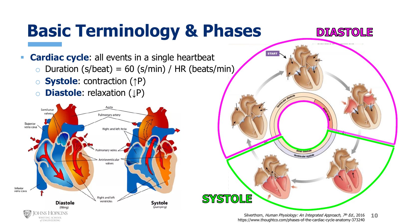
Slide 10. Ventricles filling in diastole and ejecting in systole, left, and the five stages of the cycle as a ring, right. Outer ring is ventricular events, inner ring atrial.
The cardiac cycle is all events that occur in the heart in the course of a single heartbeat. The electrical events of a single heartbeat came in the module on cardiac electrophysiology, so this lecture focuses on the mechanical, contractile events.
The duration of the cardiac cycle varies with the heart rate at the moment:
In general the cardiac cycle breaks into two phases: systole, in which the chambers contract and increase chamber pressure, and diastole, in which the chambers relax and decrease chamber pressure. In the bottom figure, the ventricles fill with blood during diastole. During systole the ventricles contract and eject that blood through their semilunar valves, into the pulmonary trunk from the right ventricle and into the aorta from the left ventricle.
There is more nuance within diastole and systole, so we break these phases into smaller stages. The figure on the right is a more typical presentation: blood entering the atria and then the ventricles through the AV valves, the atria contracting, the ventricles starting to contract, and finally the ventricles ejecting blood.
The atria and ventricles have different timing for their systole and diastole. The outer ring labels the ventricular events, with ventricular systole in the bottom two stages and ventricular diastole in the top three. The inner ring labels the atrial events, with atrial systole on the right and the other four stages atrial diastole.
Diastole also gets a proportionally bigger piece of the pie. Unless heart rate is very high, diastole simply takes up more time in the cardiac cycle.
Diastole: static and kinetic
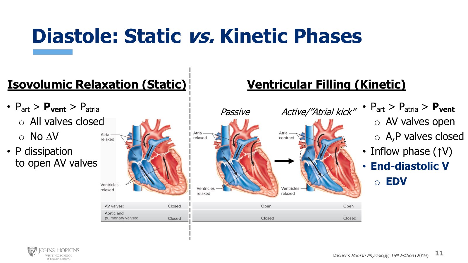
Slide 11. Isovolumic relaxation with all valves closed, then ventricular filling, passive and then active with the atrial kick. Black arrows show the pressure gradients.
Diastole has both static and kinetic elements: a stage where nothing is moving and a stage with measurable movement.
Isovolumic relaxation (static). The first stage of diastole occurs immediately after the heart has stopped contracting. Iso is a prefix meaning the same, and volumic refers to the volume of blood within the ventricle. In this phase no blood is moving, and the volume of blood in the ventricles stays the same throughout.
No blood moves because, as the ventricle starts to relax, ventricular pressure has dropped below arterial pressure on the other side of the semilunar valve, yet it is still higher than atrial pressure.
The pressure gradients close every valve in the heart. With neither inlet nor outlet open, no blood can enter or leave the ventricles: isovolumic. The black arrows on the figure show the direction of the pressure gradients. High arterial pressures close the aortic and pulmonary valves, and medium ventricular pressures close the AV valves.
The purpose of a stage with no change in ventricular blood volume: it is a transitory, temporary stage that lets the heart relax enough for the ventricular pressure generated during contraction to completely dissipate.
Ventricular filling (kinetic). Once the ventricles have relaxed enough, their pressure falls below atrial pressure. The AV valves open, and ventricular filling begins.
The aortic and pulmonary valves are still closed because ventricular pressure is much lower than arterial pressure. The inflow of blood accumulates in the ventricles, filling them more and more.
Passive. Blood simply flows from the atria into the ventricles. We spend no energy filling the ventricle; blood just pours in as it returns to the heart.
Active, the atrial kick. At some point the ventricles get full enough that the walls fight back a little, resisting further expansion, so pushing more volume in takes some force. Atrial contraction actively, forcibly squeezes blood into the ventricles, topping off the tank. This is the atrial kick, the critical final step in maximizing the volume of blood that fills the ventricle during diastole.
Once the atria have contracted and the ventricles are as full as they are going to be, the volume of blood in the ventricles is the end-diastolic volume, or EDV. The heart is now ready for ventricular contraction, or systole.
Systole: static and kinetic
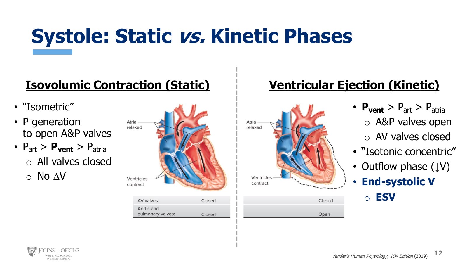
Slide 12. Isovolumic contraction with all valves closed, then ventricular ejection with the aortic and pulmonary valves open.
Systole is also divided into a static and a kinetic stage.
Isovolumic contraction (static). In this first stage no blood is moving yet, and the volume in the ventricles once again stays the same throughout. You can think of it as similar to the isometric contractions of skeletal muscle: cross-bridges form and force is generated, but muscle length does not change yet.
Muscle length does not change because the purpose of the contraction right now is to generate lots of pressure, enough ventricular pressure to overcome arterial pressure and open the aortic and pulmonary valves. The ventricle bears down on itself with all its squeezing force going toward raising ventricular pressure, and generating that kind of pressure takes a little time.
Ventricular pressure is higher than atrial pressure, which closes the AV valves, but still lower than arterial pressure, which keeps the aortic and pulmonary valves closed. With all valves closed, no blood moves anywhere in the heart, and ventricular volume does not change.
Ventricular ejection (kinetic). Once the ventricles finally generate enough pressure to overcome arterial pressure, the second stage of systole begins.
Ventricular pressure above arterial pressure opens the aortic and pulmonary valves, letting blood eject into the aorta and pulmonary trunk. Ventricular pressure much higher than atrial pressure keeps the AV valves closed.
Ejection is a little like the isotonic concentric contractions of skeletal muscle: cross-bridges form, force is produced, and the muscle shortens. When muscle length shortens in the heart, the chamber gets smaller and blood is forced out, giving ventricular outflow and a decrease in ventricular blood volume.
By the end of the ejection phase, which is also the end of systole overall, the residual blood left in the ventricles is the end-systolic volume, or ESV. There will always be some residual blood left over after contraction; the heart is designed to work this way. But end-systolic volume is variable and fluctuates with several factors covered in lecture 7F.
Summary
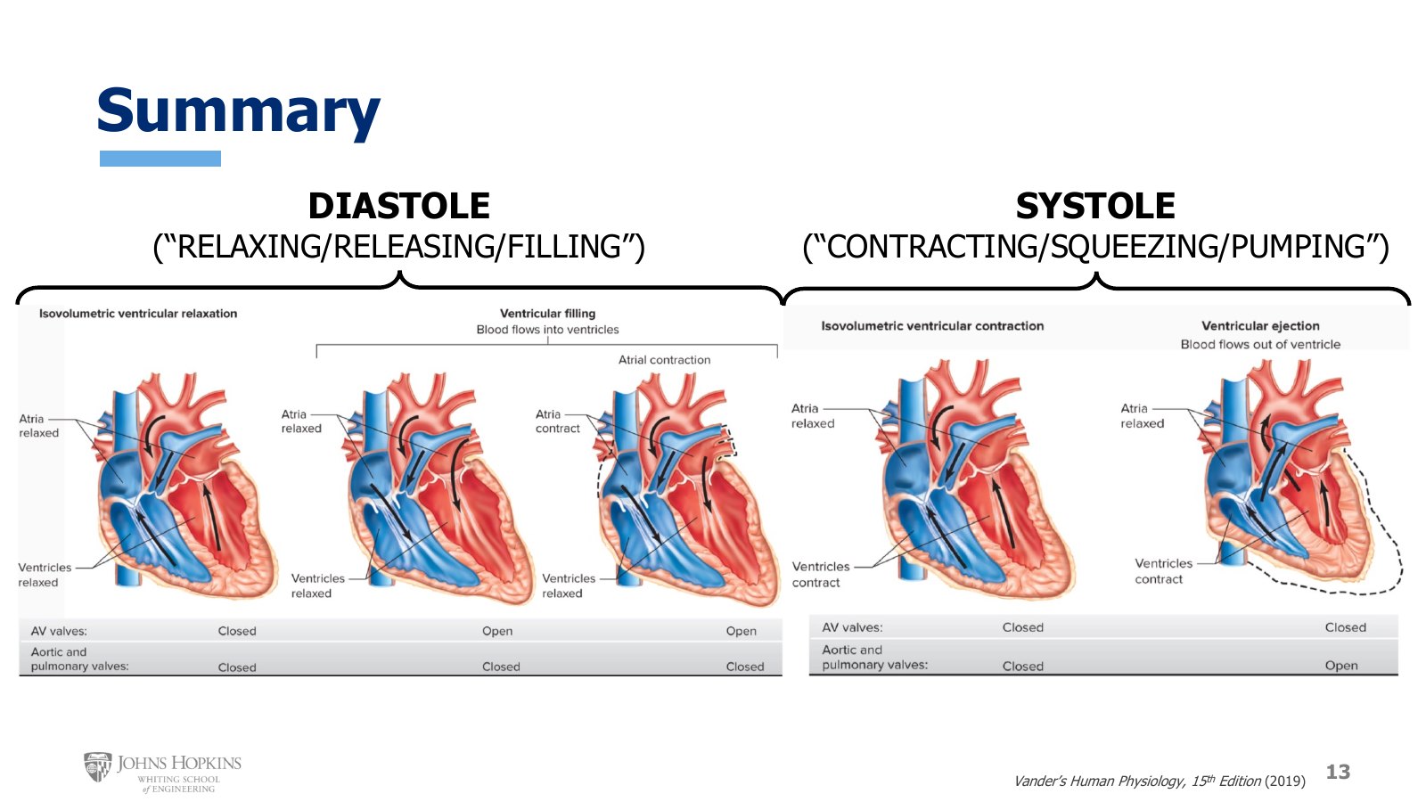
Slide 13. The three stages of diastole and two of systole in sequence, with the state of the AV and semilunar valves under each.
The summary brings the three stages of diastole and the two stages of systole together into one sequence, showing how the two previous slides work together to cover all events of a single cardiac cycle.
The five stages of the cardiac cycle
Stage | Phase | Pressures | AV valves | Aortic and pulmonary | Volume |
|---|---|---|---|---|---|
Isovolumic relaxation | Diastole, static | Part > Pvent > Patria | Closed | Closed | No change |
Passive filling | Diastole, kinetic | Part > Patria > Pvent | Open | Closed | ↑ |
Atrial kick | Diastole, kinetic | Part > Patria > Pvent | Open | Closed | ↑ to EDV |
Isovolumic contraction | Systole, static | Part > Pvent > Patria | Closed | Closed | No change |
Ventricular ejection | Systole, kinetic | Pvent > Part > Patria | Closed | Open | ↓ to ESV |