9A · Autonomic Innervation of the Cardiovascular System
This lecture reviews and clarifies how the various structures of the cardiovascular system are innervated and controlled by the autonomic nervous system. We have discussed bits and pieces of autonomic innervation at different points this semester. Here all of that information, plus a little new information, comes together on one slide to create a big picture of how the autonomic nervous system works overall.
The autonomic nervous system in general
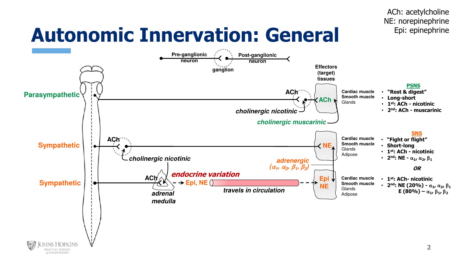
Slide 2. Parasympathetic, sympathetic, and the sympathetic endocrine variation through the adrenal medulla. The transmitter and receptor at each synapse are labeled, with the 20% norepinephrine and 80% epinephrine split for the hormone route.
On the left of the figure is a bare-bones brainstem and spinal cord. On the right are some very generic-looking target tissues, or effectors, somewhere in the body: the tissues the autonomic nerves will target.
Autonomic motor pathways in general consist of two neurons in sequence. A preganglionic neuron originates in the spinal cord and synapses on a ganglion, a cluster of neuron cell bodies somewhere in the periphery of the body. A postganglionic neuron then originates in the ganglion, travels out to the target tissue, and synapses on it.
Parasympathetic. Many of its nerves project out of the brainstem, and also out of the pelvic region of the spinal cord, although that is not pictured. Its slogan is rest and digest. The preganglionic neuron is very long, the ganglion is typically far out in the body pretty close to the target tissue, and the postganglionic neuron is quite short. Target tissues include cardiac muscle, smooth muscle, and various glands, with cardiac and smooth muscle bolded because they are of particular interest right now.
Acetylcholine released from the preganglionic neuron binds a cholinergic nicotinic receptor on the postganglionic neuron. Cholinergic just means related to acetylcholine. The postganglionic neuron also releases acetylcholine, but this acetylcholine binds cholinergic muscarinic receptors on the target tissue.
Sympathetic. The traditional sympathetic pathway has neurons projecting out of the chest and abdominal regions of the spinal cord. Its slogan is fight or flight. The preganglionic neuron is typically quite short, the ganglion is typically found pretty close to the spinal cord, and the postganglionic neuron is long. Target tissues include cardiac muscle, smooth muscle, various glands, and adipose, or fat, tissue.
Acetylcholine from the preganglionic neuron again binds a cholinergic nicotinic receptor on the postganglionic neuron. The postganglionic synapse at the tissues is where everything changes. No more acetylcholine release or cholinergic receptors. Postganglionic sympathetic neurons release norepinephrine, which binds adrenergic receptors. Adrenergic receptors come in four major subtypes: alpha-1, alpha-2, beta-1, and beta-2. Norepinephrine likes to bind alpha-1, alpha-2, and beta-1 receptors.
The endocrine variation. A variation on the sympathetic pathway reinforces the neural sympathetic effects on the body. A sympathetic preganglionic neuron still emerges from the spinal cord, but it does not synapse at a ganglion. Instead it synapses on the central region of the adrenal gland, called the adrenal medulla. Its acetylcholine binds cholinergic nicotinic receptors on adrenal medulla cells, which stimulates the adrenal medulla to release epinephrine and norepinephrine into the bloodstream. That makes them hormones now.
Epinephrine and norepinephrine are chemical and physiological cousins. They circulate around the body, exit at tissue capillaries, and bind any tissue that has an adrenergic receptor, which is still cardiac muscle, smooth muscle, glands, and adipose.
Hormone norepinephrine, about 20% of the hormone release, is the exact same chemical released from sympathetic nerve endings, so it uses the exact same receptors: alpha-1, alpha-2, and beta-1.
Epinephrine, the remaining 80% of hormone release, likes to bind alpha-1, beta-1, and beta-2 receptors.
The three autonomic routes to an effector
Route | First neuron | Ganglion | At the ganglion | At the target |
|---|---|---|---|---|
Parasympathetic | Long | Far out, near the target | ACh on nicotinic | ACh on muscarinic |
Sympathetic, neural | Short | Close to the spinal cord | ACh on nicotinic | Norepinephrine on alpha-1, alpha-2, beta-1 |
Sympathetic, endocrine | Goes to the adrenal medulla | None | ACh on nicotinic, adrenal medulla | Norepinephrine (20%) on alpha-1, alpha-2, beta-1. Epinephrine (80%) on alpha-1, beta-1, beta-2 |
Autonomic innervation of cardiovascular structures
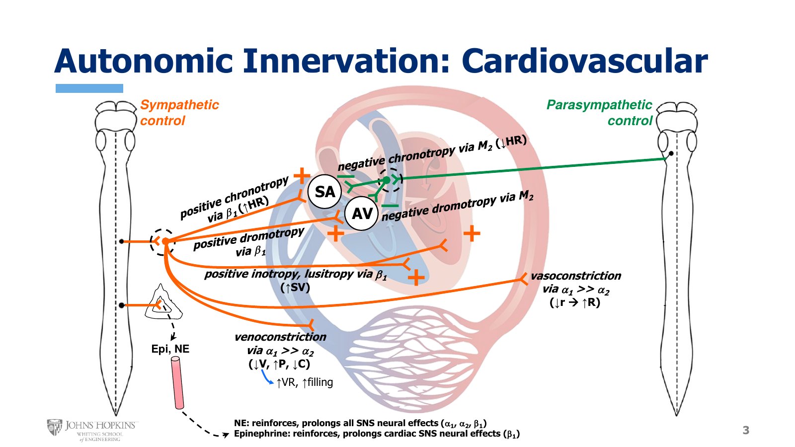
Slide 3. Sympathetic control on the left, parasympathetic on the right. Plus signs mark sympathetic effects at the SA node, AV node, myocardium, arteries, and veins. Parasympathetic fibers reach only the SA and AV nodes through M₂ receptors.
Narrowing the focus to cardiovascular structures, sympathetic innervation is mapped on the left, emerging from the spinal cord and traveling out to the heart and blood vessels, and parasympathetic innervation on the right. Both types of nerves exist on both sides of the body. They are separated here only for clarity. Working from top to bottom:
SA and AV nodes. As we know from the cardiac electrophysiology module, when sympathetic norepinephrine binds beta-1 receptors at the sinoatrial node, the result is a positive chronotropic effect, otherwise known as an increase in heart rate. When sympathetic norepinephrine binds beta-1 receptors at the atrioventricular node, the result is a positive dromotropic effect: conduction velocity through the AV node increases, which shortens the delay of the action potential.
Parasympathetic nerves also innervate the SA and AV nodes, and they exert the opposite effect. When acetylcholine binds muscarinic-2, or M2, receptors on those structures, parasympathetic stimulation produces negative chronotropy, a slower heart rate, at the SA node, and negative dromotropy, slower conduction velocity, through the AV node. So far this is a balanced dual innervation, with sympathetic and parasympathetic nerves exerting equal and opposite influences over cardiac electrical activity.
Myocardium. Innervation of the myocardium, the cardiac muscle, is mostly just sympathetic. When norepinephrine binds beta-1 receptors on the myocardium, it changes intracellular calcium levels so that cardiac muscle cells contract more forcefully and relax more completely. That is positive inotropy and positive lusitropy, which together contribute to a higher stroke volume.
Arteries. Innervation of the blood vessels, arteries and veins both, is also mostly sympathetic. On the arterial side, sympathetic norepinephrine binds alpha receptors, with alpha-1 receptors outnumbering alpha-2 receptors in most of the vasculature. This contracts the arterial vascular smooth muscle: vasoconstriction. Constriction gives a smaller internal radius, and so a higher vascular resistance.
Veins. Sympathetic stimulation of the veins produces a similar contraction of vascular smooth muscle, but on the venous side of the circulation it is called venoconstriction. When the veins constrict, venous volume decreases as the constriction forces venous blood back toward the heart, venous pressure increases, and overall venous compliance decreases. Because that contraction forces venous blood back toward the heart, venoconstriction also increases venous return, which increases ventricular filling and so increases preload.
The endocrine side. In the name of full disclosure, the sympathetic endocrine side is engaged here too, releasing mostly epinephrine and some norepinephrine into the bloodstream. They circulate around the body and reinforce and prolong what the sympathetic nerves have already been doing.
Autonomic control of blood pressure
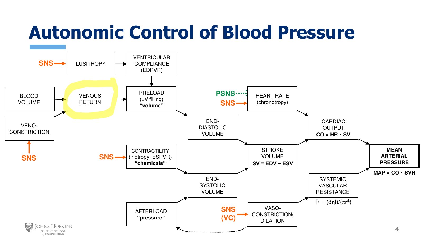
Slide 4. The determinants of cardiac output flowchart, now extended to mean arterial pressure. Orange SNS arrows and a dotted green PSNS arrow mark where the autonomic nerves act. Venous return is highlighted for later in the module.
Earlier this semester, when we talked about determinants of cardiac output, we introduced a flowchart. At that point we had only discussed the cardiac elements of the system, no vessels yet, so it was limited to heart stuff:
Cardiac output, which equals heart rate times stroke volume.
Heart rate.
Stroke volume, which equals end-diastolic volume minus end-systolic volume.
Preload, related to ventricular filling volume.
Contractility, related to chemicals that change calcium availability in cardiac muscle cells.
Afterload, related to the pressures found in both the arteries and the ventricle.
We have learned a lot since then, so now we can fill in more of the big cardiovascular picture. Cardiac output is a determinant of mean arterial pressure, along with systemic vascular resistance.
The rearranged version of the Hagen-Poiseuille equation solves for vascular resistance: eight times blood viscosity times vessel length, all divided by π times radius raised to the fourth power.
Radius is bolded because we have dedicated vasoconstrictor and vasodilator mechanisms that manipulate radius to regulate vascular resistance and arterial pressure. Because of their role in altering resistance and pressure, vasoconstriction and vasodilation also become primary determinants of cardiac afterload.
We have also learned more about factors that affect preload, or ventricular filling.
Ventricular compliance. A more compliant ventricular wall can stretch and accommodate more filling during diastole. The term typically refers to permanent mechanical wall tissue changes that stiffen the ventricular wall in conditions like aging, diabetes, and heart disease.
Lusitropy. The ability of the heart to relax quickly and completely can also affect filling and compliance.
Venous return. A vascular factor. Venous return, the flow rate of blood from the veins back into the heart, supplies the filling volume for the ventricles. Both overall blood volume in the body and the degree of venoconstriction directly impact venous return. High venous return gives high preload, and low venous return gives low preload.
What does all this have to do with neurological innervation? The flowchart shows the effects sympathetic and parasympathetic stimulation have on cardiovascular function, and so how autonomic nervous system activity regulates arterial pressure. The focus is arterial pressure because arterial blood pressure is a major variable our cardiovascular reflexes are gauged to monitor and regulate.
Sympathetic stimulation increases blood pressure. It increases heart rate, enhances lusitropy, promotes venous return and ventricular filling by stimulating venoconstriction, increases contractility, and stimulates arterial vasoconstriction.
Parasympathetic stimulation decreases blood pressure by decreasing heart rate. Along with parasympathetic stimulation there is also a withdrawal of sympathetic stimulation, and so a loss of the major sympathetic effects.