9C · Elements of the Atrial Stretch Reflex: The Bainbridge Reflex
The atrial stretch reflex has both neural and endocrine subcomponents. This lecture focuses on its major neural subcomponent, the Bainbridge reflex. If the baroreflex is how we monitor and respond to changes in blood pressure, the atrial stretch reflex, a huge umbrella term for several smaller related reflex loops, is how we monitor and respond to changes in blood volume. Once again negative feedback keeps blood volume from fluctuating outside its normal homeostatic range.
The entire atrial stretch reflex
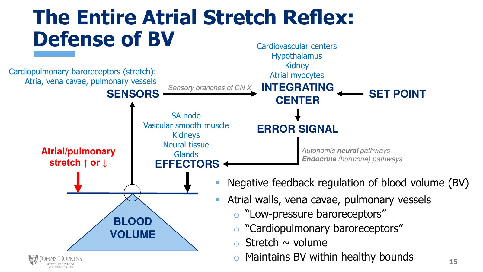
Slide 15. The negative feedback schematic with blood volume as the regulated variable. Cardiopulmonary sensors, four integrating centers, and both neural and endocrine outputs to five kinds of effector.
For the entire atrial stretch reflex, blood volume is the regulated variable, with the seesaw on top. The perturbation is when the degree of stretch in the atrial chambers of the heart and in the pulmonary circulation increases or decreases beyond acceptable bounds.
Sensors. When blood volume increases or decreases too much, it is sensed by low-pressure baroreceptors, also called cardiopulmonary baroreceptors. These are in the walls of the atria of the heart, in the vena cavae where they empty into the right atrium, and in the vessels of the pulmonary circulation.
Afferent path. These low-pressure cardiopulmonary baroreceptors send information about heart and vessel stretch, and so volume, along sensory pathways in branches of cranial nerve X, the vagus nerve.
Integrating centers. Because so many smaller reflexes make up the overall atrial stretch reflex, many different tissues receive this information and compare it with a predetermined set point:
the cardiovascular centers in the medulla
the hypothalamus, in a different part of the brain
the kidneys
the atrial myocytes, the cardiac muscle cells in the atrial chambers of the heart
Efferent paths and effectors. After an error signal is generated, the reflex tries to fix the discrepancy between actual and intended blood volume by sending outgoing messages through autonomic neural pathways and through endocrine, or hormonal, pathways. These signals reach target tissues that manipulate blood volume, and whatever pressure issues have arisen from the abnormal volume. The wide variety of target tissues includes the SA node of the heart, vascular smooth muscle in the blood vessels, the kidneys, neural tissue, and glandular tissue.
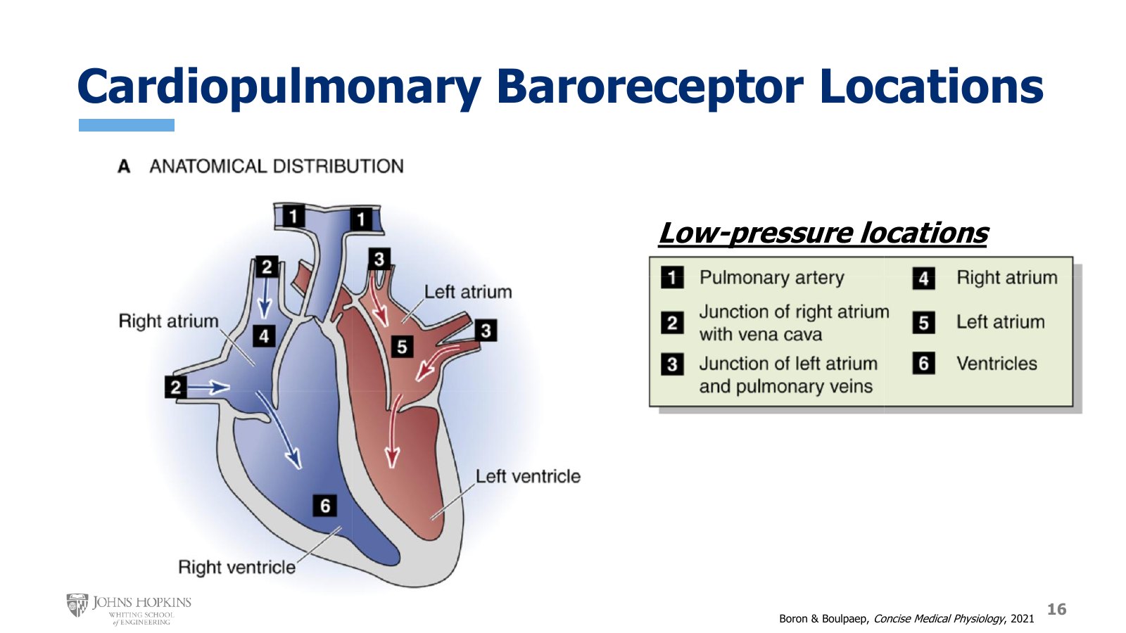
Slide 16. The six low-pressure baroreceptor locations, numbered on a heart: pulmonary artery, vena cava junction, pulmonary vein junction, right atrium, left atrium, and ventricle (marked in the right ventricle).
Stretch in these areas, where pressure is low and so not as much of a concern, is interpreted by the body as an indicator of volume: how much blood is contained in those spaces at the moment. Low-pressure baroreceptors are found in:
the pulmonary arteries
the junction where the superior and inferior vena cavae join the right atrium
the pulmonary veins as they empty into the left atrium
the right atrium itself
the left atrium itself
the right ventricle
The right ventricle seems to stick out, since it is clearly not an atrium and this is the atrial stretch reflex. But it is still a low-pressure chamber of the heart, so it can send more information about stretch and volume.
Everything so far addresses the entire atrial stretch reflex as a very broad phenomenon with both neural and endocrine elements regulating blood volume. Lecture 9D covers the endocrine elements. The rest of this lecture covers the Bainbridge reflex.
The Bainbridge reflex
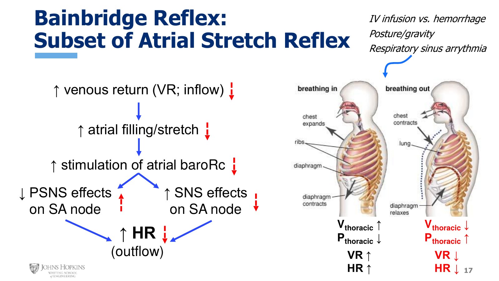
Slide 17. Left, the Bainbridge chain from venous return to heart rate, with red dashed arrows for the reverse case. Right, breathing in raises venous return and heart rate, and breathing out lowers both.
The Bainbridge reflex is a subset of the overall atrial stretch reflex. It is mainly concerned with regulating the volume of blood filling the chambers of the heart, manipulating heart rate to match the rate of outflow, cardiac output, to the rate of inflow, venous return. Pump inflow needs to match pump outflow or we start to run into problems, as lecture 9E shows. Coupling outflow with inflow maintains a steady state at the heart.
The Bainbridge reflex kicks in when venous return increases for some reason:
The rate of inflow into the heart increases.
Atrial wall stretch increases as the atria fill faster with the greater inflow.
The increased stretch stimulates the low-pressure baroreceptors in the atrial walls.
They send the message about increased stretch to the cardiovascular centers in the medulla.
The medulla sends out two complementary neural signals to deal with the volume load: decreased parasympathetic stimulation of the SA node and increased sympathetic stimulation of the SA node.
Together these increase heart rate, which empties the heart chambers more often.
We increase our outflow rate to better match the higher inflow rate as venous return increases. That prevents the chambers of the heart from filling beyond a volume they can comfortably handle.
The opposite is also true. A decrease in venous return gives less atrial stretch and less stimulation of the atrial low-pressure baroreceptors. That removes the suppression of parasympathetic signals to the SA node and inhibits sympathetic outflow to the SA node, both decreasing heart rate.
What changes venous return
A true change in blood volume. This absolutely affects venous return flow rates. Intravenous infusion of extra fluids increases it, and blood loss of some kind decreases it.
A change in blood distribution within the body. Standing up means more blood remains in the compliant veins of the legs, since gravity likes to pull blood down, so less blood returns to the heart: lower venous return. Lying down means gravity is no longer keeping blood in the legs and feet, so it is much easier for blood to return to the heart: greater venous return. If you ever land a spot to travel into space, microgravity does much the same thing as lying down, so venous return increases.
Breathing. The act of merely breathing alters venous return.
There is a detectable fluctuation in heart rate that coincides with the cycle of inhalation and exhalation, called respiratory sinus arrhythmia. Despite the scary-sounding name, it just describes the predictable oscillations in heart rate as we breathe in and out. More on breathing comes later in the semester.
Breathing in. The volume of the chest cavity, or thoracic cavity, expands and increases. Through an application of Boyle’s law, the pressure inside the thoracic cavity decreases. We are essentially creating a negative, subatmospheric pressure in there, a vacuum, which is what literally sucks air from the atmosphere into the lungs. That vacuum does not just suck air into the chest. It also sucks blood into the chest, increasing venous return through the vena cava vessels into the right atrium. The consequent increase in right atrial stretch triggers the Bainbridge reflex, increasing heart rate every time we breathe in.
Breathing out. The opposite events unfold. The thoracic cavity contracts inward and its volume decreases, so the pressure inside increases. Thoracic pressure is now higher than atmospheric pressure, which is what literally pushes air out of the lungs. That positive pressure also discourages venous return to the right atrium. The decrease in right atrial stretch stops triggering the Bainbridge reflex, decreasing heart rate every time we breathe out.
The baroreflex and the Bainbridge reflex together
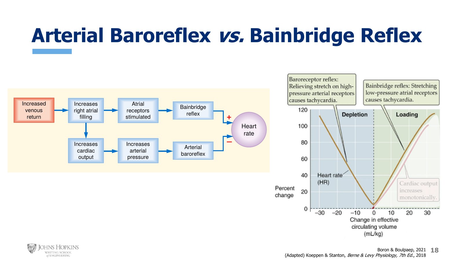
Slide 18. Left, one stimulus drives both reflexes with opposite effects on heart rate. Right, percent change in heart rate against change in effective circulating volume (mL/kg). The V-shaped curve is labeled baroreceptor reflex on the depletion side and Bainbridge reflex on the loading side.
Both reflexes monitor hemodynamic variables near the heart all the time, so consider what happens when they are engaged at the same time. Start with something that increases venous return: receiving intravenous fluids, lying down, or simply breathing in. The high venous return increases right atrial filling.
Through the baroreflex. Right atrial filling and pressure is a very upstream surrogate of preload. Higher right atrial filling means higher stroke volume through the Frank-Starling mechanism, and so higher cardiac output. Higher cardiac output gives higher blood pressure, which stimulates the baroreflex and, among other things, produces a reflex decrease in heart rate.
Through the Bainbridge reflex. The same increase in right atrial filling stimulates the atrial stretch receptors, engaging the Bainbridge reflex and producing a reflex increase in heart rate.
The same stimulus engages the two major neurological cardiovascular reflexes, but they respond in opposite ways, because each prioritizes a different aspect of hemodynamic stability and homeostasis. The baroreflex defends arterial pressure, which here requires a decrease in heart rate. The Bainbridge reflex defends atrial filling volume, which here requires an increase in heart rate.
There is a logic to how the two give and take, with one dominating the other depending on the physiological circumstances. The graph plots percent change in heart rate on the y-axis against change in circulating blood volume on the x-axis. The heart rate curve looks like a V: high values at very low and at very high blood volumes, and its baseline lowest value at normal circulating blood volume, neither excessively high nor excessively low.
The baroreflex dominates when blood volume falls below baseline. Decreased volume decreases heart filling and cardiac output, and so arterial pressure. Low arterial pressure increases sympathetic outflow to the SA node, raising heart rate. The rise in heart rate that accompanies decreases in circulating volume occurs through the baroreflex.
The Bainbridge reflex dominates when blood volume rises above baseline. Increased circulating volume increases atrial filling and atrial stretch, and so heart rate.