9B · The Baroreflex
The arterial baroreflex is a very well-known and well-studied cardiovascular reflex. It is our major regulator of arterial blood pressure, keeping it within healthy homeostatic bounds. If pressure suddenly gets too high, we risk rupturing vessels that are not designed to withstand those pressures. If it suddenly gets too low, we sacrifice brain perfusion, because we no longer have the driving pressure for cerebral blood flow. So negative feedback mechanisms keep mean arterial pressure on an even keel and prevent it from fluctuating too much in either direction.
The baroreflex as a negative feedback loop
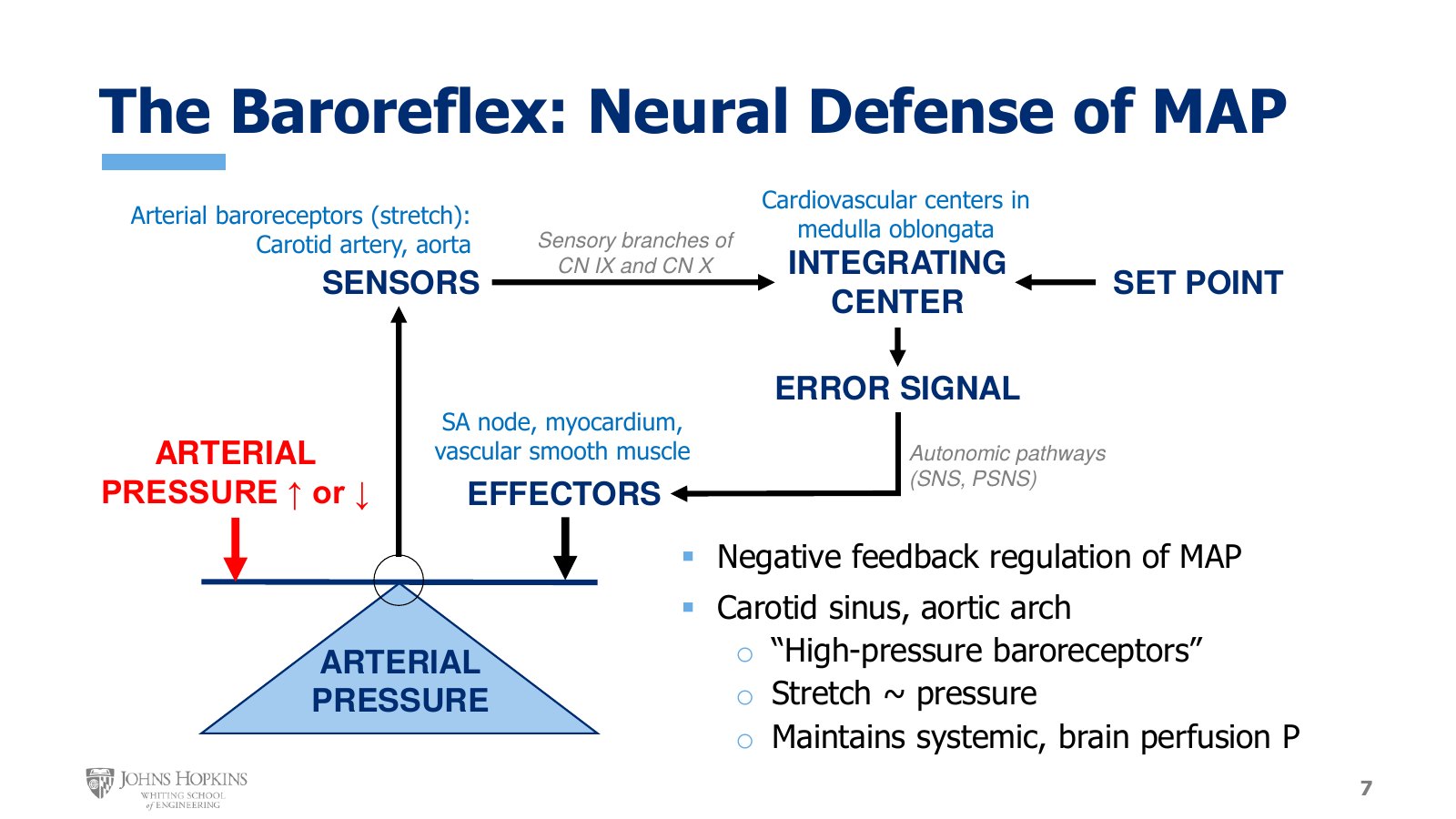
Slide 7. The Module 1 negative feedback schematic with arterial pressure as the regulated variable. Sensors in the carotid artery and aorta, integration in the medulla oblongata, and autonomic output to the SA node, myocardium, and vascular smooth muscle.
This is the negative feedback schematic introduced at the beginning of the semester. Arterial pressure is the regulated variable, with a seesaw on top of it to symbolize the balance and equilibrium we need to establish and maintain. The red down arrow on the left is a perturbation that throws the seesaw out of balance. In this case, arterial pressure acutely increases or decreases beyond acceptable bounds. The rest of the diagram works toward pushing down on the black arrow on the right to restore balance to the system.
Sensors. A sudden increase or decrease in arterial pressure is sensed by baroreceptors in two major arteries: the carotid artery, monitoring blood pressure in vessels feeding the brain, and the aorta, monitoring blood pressure in the major vessel feeding the entire systemic circulation.
Afferent path. Baroreceptors send information about arterial wall stretch, and so arterial blood pressure, along sensory neurons in branches of cranial nerve IX, the glossopharyngeal nerve, and cranial nerve X, the vagus nerve.
Integrating center. The sensory pressure information is integrated in the cardiovascular control centers in the medulla oblongata, the most primitive part of the brainstem. If incoming arterial pressure differs from the set point for where it needs to be, the brainstem generates an error signal and initiates motor commands out to the cardiovascular tissues of the body to fix the problem.
Efferent path and effectors. The outgoing motor signals are carried by the autonomic nerves, sympathetic and parasympathetic, to the effectors, the target cardiovascular tissues that will fix the problem: the SA node of the heart, the myocardium, and vascular smooth muscle.
For a big-picture recap: arterial baroreceptors are found in the carotid artery, specifically the carotid sinus, where the common carotid splits into the internal and external carotid arteries, and in the aorta, specifically the aortic arch. They are often called high-pressure baroreceptors because they sit in the large arteries of the systemic circulation, the highest-pressure blood vessels we have. Stretch in the walls of high-pressure arteries is interpreted as an index of pressure, and the baroreflex exists to maintain perfusion pressure, ensuring adequate blood flow to the brain and the systemic circulation.
Where the arterial baroreceptors are
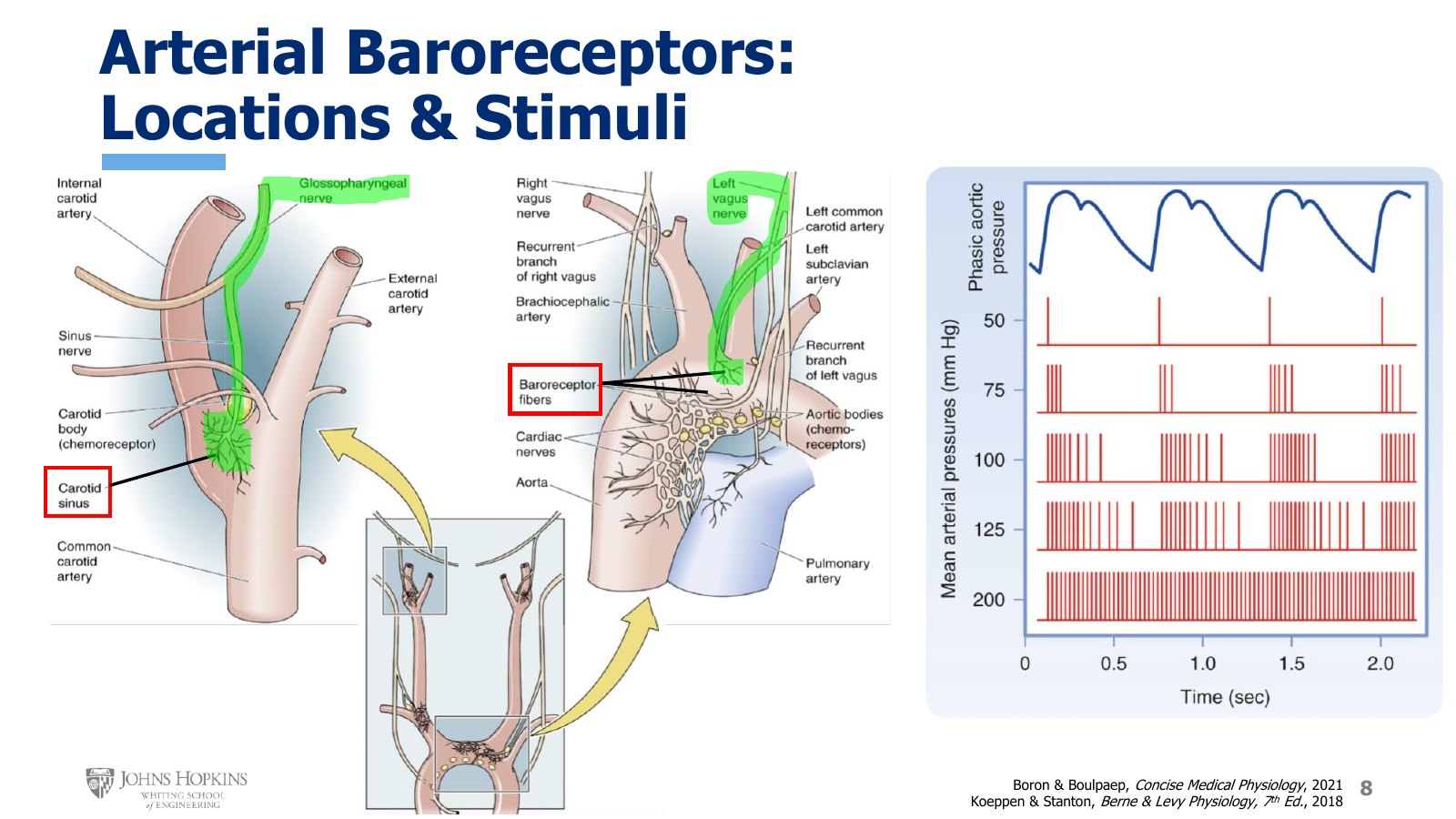
Slide 8. Left, the carotid sinus with its glossopharyngeal branch highlighted. Center, aortic arch baroreceptor fibers on the left vagus. Right, baroreceptor firing under a phasic aortic pressure trace at mean arterial pressures of 50, 75, 100, 125, and 200 mmHg.
The figure shows the ascending aorta and the aortic arch, with the common carotid arteries branching off and heading up toward the brain on the right and left sides of the body. Zooming in gives a better sense of receptor location and innervation.
Carotid. The common carotid artery comes up and splits into the external carotid artery, which feeds the scalp and face, and the internal carotid artery, which feeds the brain. A slight swelling at the base of the internal carotid is the carotid sinus, where the carotid artery baroreceptors are found. These receptors are innervated by a branch of the glossopharyngeal nerve. That nerve projects out of the brainstem, so it delivers information about carotid stretch and pressure directly to the cardiovascular centers in the brainstem.
Aortic. The aortic receptors are concentrated in the arch of the aorta, before it turns and heads down through the chest and abdomen. They are innervated by a branch of the vagus nerve, another nerve that projects out of the brainstem and so delivers aortic stretch and pressure information straight to the cardiovascular centers there.
Firing rate tracks pressure
At the top of the right panel is an arterial pressure waveform. It could be aortic or carotid, and it is intended to be generic. Systolic pressure peaks at the top, then pressure subsides during diastole until it reaches its lowest diastolic value right before the next systole starts. There is an x-axis label of time, but deliberately no y-axis pressure values for the waveform.
Moving down the figure are the patterns of baroreceptor firing bursts when mean arterial pressure is extremely low, pretty low, fairly average, moderately high, and extremely high.
Extremely low. The baroreceptors fire only at the very beginning of systole, when pressure and stretch suddenly start to increase, and that is it for the entire cardiac cycle. Virtually no baroreceptor stimulation.
A little higher, still pretty low. A few more bursts of depolarization are added during the uphill systolic phase of the pressure wave.
Rising further. Each level of pressure stimulates the baroreceptors more, adding more bursts of depolarization.
Very highest pressure. With the highest levels of arterial wall stretch, the baroreceptors fire waves of depolarization pretty much all the time in a high-frequency pattern.
Response to a too-high pressure
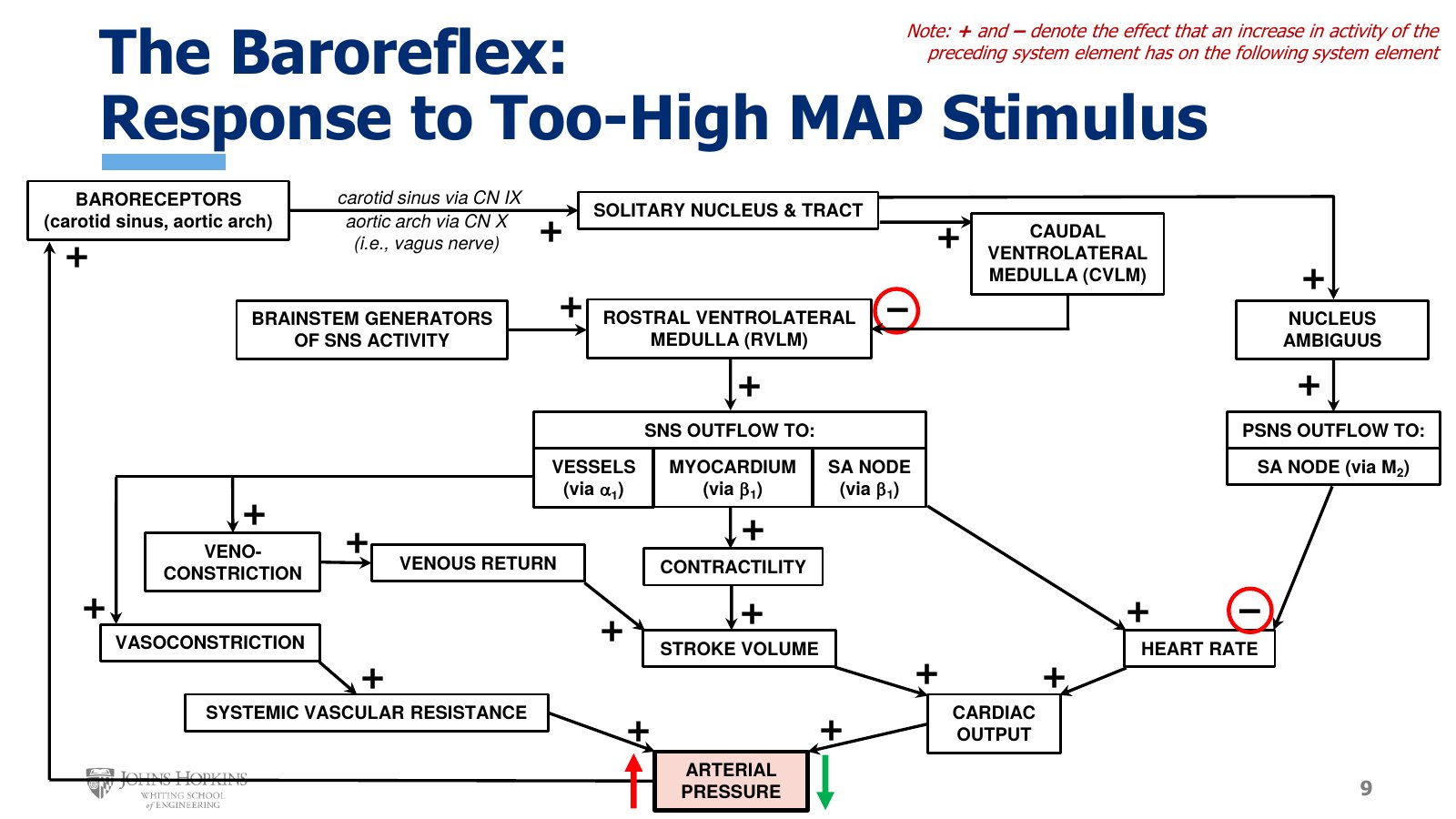
Slide 9. The baroreflex flowchart for a rise in pressure. Plus and minus signs give the effect an increase in one element has on the next. The CVLM inhibits the RVLM, and parasympathetic outflow inhibits heart rate.
The flowchart maps the order of events in the reflex from start to finish. The plus and minus signs indicate the effect that an increase in the activity of one element has on the very next element. An increase in baroreceptor firing rate has a stimulatory effect on the solitary nucleus and tract, while an increase in parasympathetic outflow to the SA node has an inhibitory effect on heart rate.
Take the scenario of arterial blood pressure that is momentarily too high, such as doing a handstand. That places the carotid artery and aortic arch below heart level and increases the pressure the baroreceptors see.
Arterial pressure, the regulated variable, increases. The baroreceptors are stimulated, depolarize more often, and increase their firing rate.
The increased stimulation is transmitted by the glossopharyngeal nerve, cranial nerve IX, and the vagus nerve, cranial nerve X. These stimulate a region of the cardiovascular centers in the medulla known as the solitary nucleus and tract.
The activated solitary nucleus and tract stimulates two other regions of the medullary cardiovascular centers: the caudal ventrolateral medulla, or CVLM, and the nucleus ambiguus.
The stimulated nucleus ambiguus stimulates parasympathetic outflow to the sinoatrial node, which has an inhibitory effect and decreases heart rate.
The activated CVLM inhibits its brainstem neighbor, the rostral ventrolateral medulla, or RVLM. The job of the RVLM is to drive sympathetic outflow to all our cardiovascular tissues.
With the RVLM inhibited, sympathetic outflow to the heart and blood vessels is inhibited. Sympathetically mediated increases in heart rate, contractility, vasoconstriction, and venoconstriction are withdrawn.
Does a decrease in heart rate make sense as a response to high blood pressure? Yes. Lower heart rate leads to lower cardiac output, which helps lower blood pressure back into a normal range. The loss of baseline sympathetic influences on cardiovascular function decreases systemic vascular resistance and further decreases cardiac output, also supporting an appropriate reflex decrease in arterial pressure back down to normal.
Response to a too-low pressure
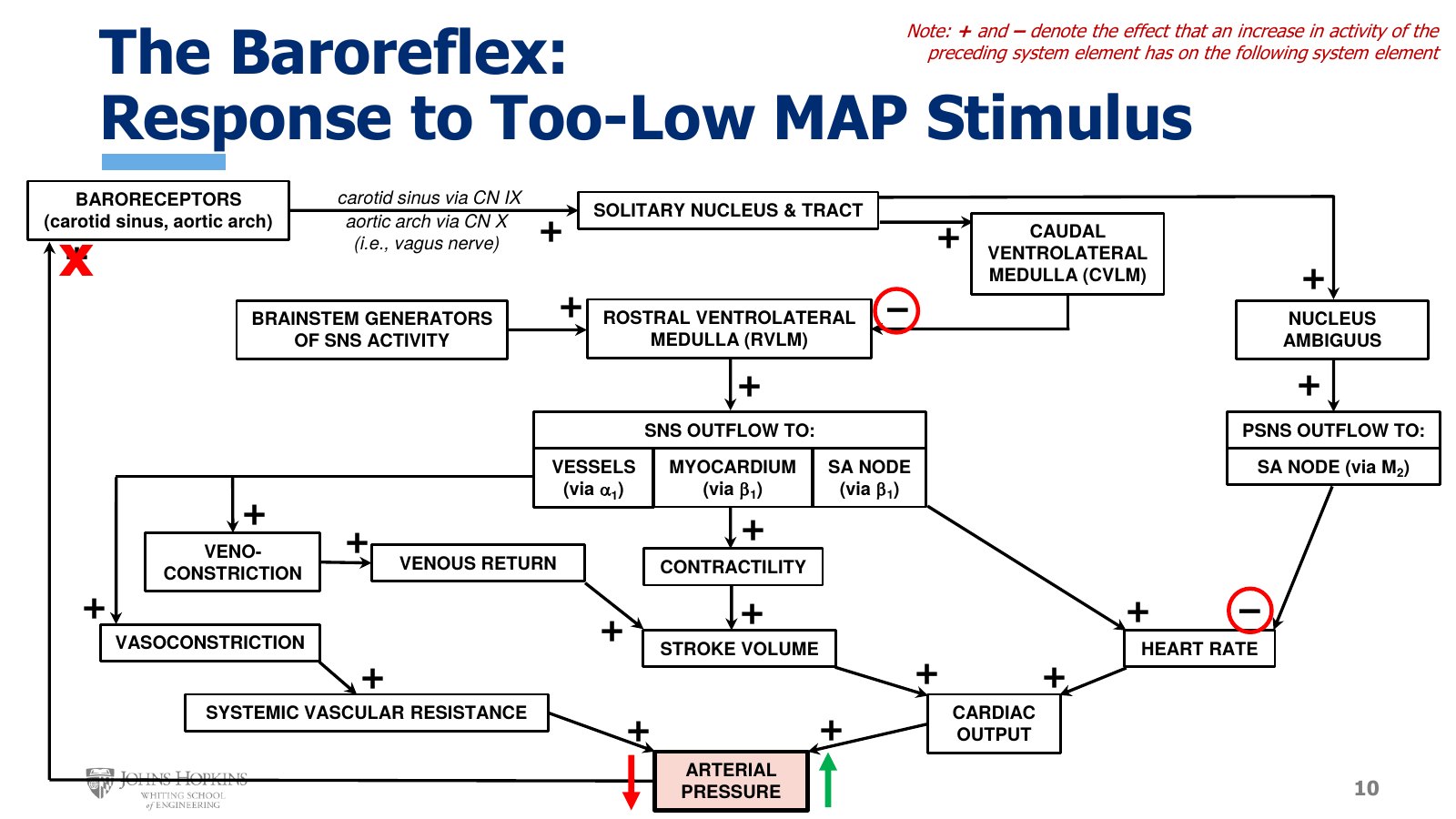
Slide 10. The same flowchart for a fall in pressure. The red X at the baroreceptors marks the loss of firing, which removes the CVLM brake on the RVLM.
Sudden increases in arterial pressure occur less often than sudden decreases, and sudden decreases threaten our health to a greater extent. A loss of arterial pressure means a loss of the pressure that drives perfusion of the brain, and a loss of brain blood flow leads to death pretty quickly. So the response to a sudden decrease is the more significant application of the baroreflex in our cardiovascular toolbox.
Arterial pressure is still the regulated variable. A decrease might come with blood loss, significant dehydration, standing in the heat for too long, or simply standing up too quickly from a sitting or lying position.
With arterial pressure too low, the baroreceptors are no longer stimulated to fire.
Without ongoing baroreceptor stimulation, every downstream effect of stimulating the baroreceptors, and so the solitary nucleus and tract, stops. Parasympathetic outflow from the nucleus ambiguus goes away, as does stimulation of the CVLM and its inhibition of the RVLM.
The RVLM, which sends the sympathetic signals to the heart and blood vessels, is no longer inhibited. You could say it is disinhibited, and it is now free to drive sympathetic outflow.
SA node: heart rate increases, so cardiac output increases.
Myocardium: contractility increases, giving higher stroke volume and so higher cardiac output.
Arteries: vasoconstriction, and a subsequent increase in systemic vascular resistance.
Veins: venoconstriction, which drives an increase in venous return, which drives higher preload, giving higher stroke volume and so higher cardiac output.
The combination of higher cardiac output and higher vascular resistance raises arterial blood pressure, restoring it to the normal range.
A 500 mL hemorrhage simulation
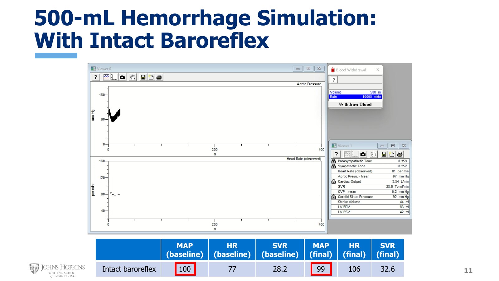
Slide 11. Simulation with the baroreflex intact. Aortic pressure on top, heart rate below, live values at right. The table gives the baseline and final MAP, heart rate, and SVR.
Computer simulations show how important the baroreflex truly is when arterial pressure drops. These simulate a 500 mL loss of blood volume, which is what we experience when we give blood, a blood loss equivalent to a class one hemorrhage.
The display has real-time chart recordings of arterial pressure on top and heart rate on the bottom. It also has real-time values for parasympathetic and sympathetic nerve activity, both on a scale of 0 to 1, with zero being no neural activity and one being maximal. Heart rate values are listed because specific values are hard to read off the chart, and mean aortic pressure values because the chart shows only the pulsatile waveform. The box also lists cardiac output, systemic vascular resistance, central venous pressure, carotid sinus pressure, and ventricular volumes. Once a stable baseline was reached, a snapshot of baseline values was taken and the blood loss was started.
Intact baroreflex. At baseline, mean aortic pressure was 100 mmHg, heart rate 77 beats per minute, and systemic vascular resistance 28.2 torr per liter per minute. One torr is approximately equal to one millimeter of mercury. After the hemorrhage, with the baroreflex free to respond to the blood loss and the fall in arterial pressure, mean aortic pressure ended at 99 mmHg, heart rate at 106 beats per minute, and systemic vascular resistance at 32.6 torr per liter per minute. The baroreflex restored arterial pressure by increasing both cardiac and vascular responses, through a decrease in parasympathetic activity and an increase in sympathetic activity.
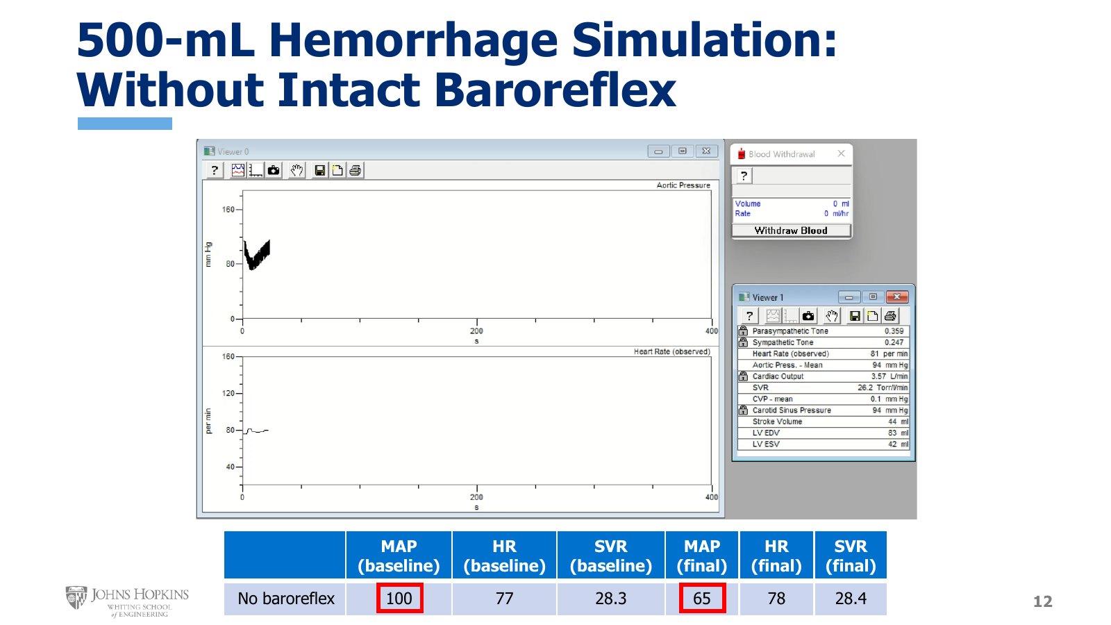
Slide 12. The same hemorrhage with autonomic outflow clamped at baseline. Heart rate and SVR barely move, and MAP falls to 65 mmHg.
No baroreflex. The second run uses the exact same scenario but prevents the parasympathetic and sympathetic nervous systems from responding to the drop in pressure. This disables the efferent, or motor, or outgoing arm of the reflex that communicates with the cardiovascular tissues. Clicking the locks next to the parasympathetic and sympathetic outflow data unlocks those variables from the simulation, so they can no longer respond. The nervous system activity seen at baseline stays clamped for the entire hemorrhage.
As the hemorrhage proceeds, heart rate does not change, and neither does systemic vascular resistance, because both depend on autonomic input and both types of autonomic nerves are clamped. Nothing is changing. At baseline, mean aortic pressure was 100 mmHg, heart rate 77 beats per minute, and systemic vascular resistance 28.3 torr per liter per minute. After the hemorrhage, heart rate remained at 78 beats per minute and systemic vascular resistance at 28.4. Without either compensatory mechanism, mean arterial pressure fell to 65 mmHg, low enough to make most people pass out from lack of brain blood flow.
500 mL hemorrhage, baseline to final
Condition | MAP (mmHg) | Heart rate (beats/min) | SVR (torr/L/min) |
|---|---|---|---|
Intact baroreflex | 100 → 99 | 77 → 106 | 28.2 → 32.6 |
No baroreflex | 100 → 65 | 77 → 78 | 28.3 → 28.4 |