9D · Elements of the Atrial Stretch Reflex: Endocrine Reflexes
The endocrine aspects of the atrial stretch reflex use hormones instead of neural transmission to carry negative feedback regulatory messages to our tissues. Neural function has been the primary focus of this module so far. Zooming out shows the different forms of negative feedback communication in the bigger picture.
Neural versus endocrine reflexes
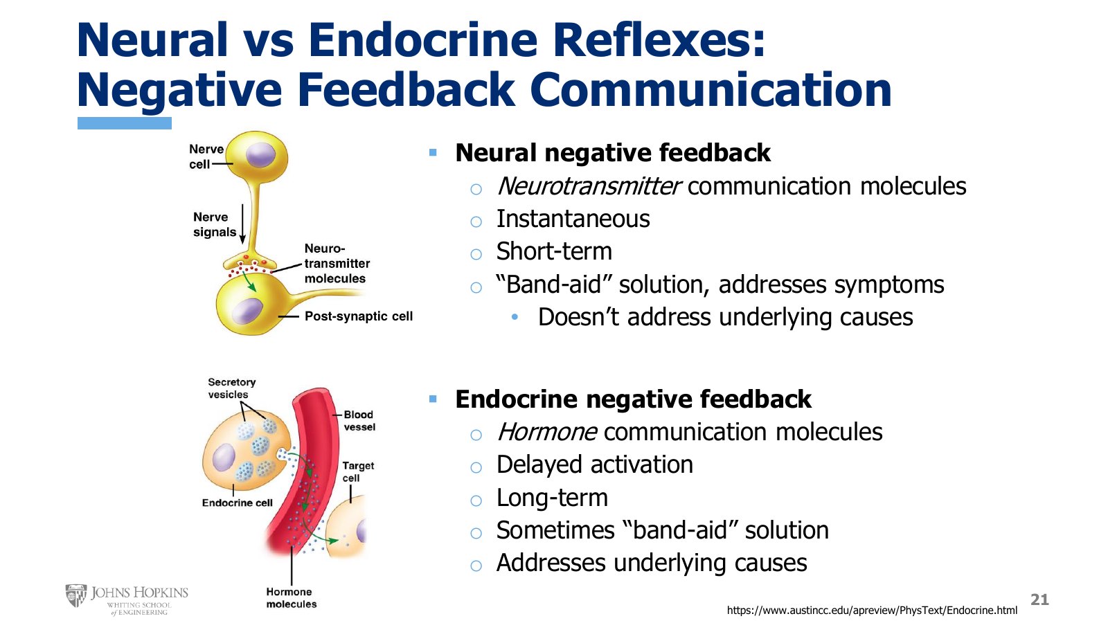
Slide 21. Neurotransmitter crossing a synapse above, hormone traveling through a blood vessel to a target cell below, each with its feedback properties listed.
Neural reflexes rely on messenger molecules released from a neuron that diffuse across a tiny synaptic cleft and bind receptors on the postsynaptic cell membrane, changing the behavior of that postsynaptic cell. In neural reflexes these chemical messengers are called neurotransmitters.
Virtually instantaneous. Nerves conduct electrical signals so quickly that they can fix a regulatory problem in the blink of an eye.
Short-lived. The flip side of being fast to turn on is being fast to turn off. Nerve cells cannot keep up that level of nonstop corrective messaging forever. Neurons can experience fatigue.
A band-aid solution. Neural reflexes are great at fixing a short-term departure from homeostasis, but they usually address the problematic symptoms of the imbalance and often not the underlying issues that created it.
Neural reflexes are lightning fast, keep us alive from second to second, and are absolutely essential for survival. We also need something that acts more long-term and can address the underlying root causes of a physiological imbalance. For that we rely on endocrine reflexes.
Endocrine reflexes use chemical messengers called hormones. Hormones are released from a gland of some sort, enter the bloodstream, and circulate around the body until they arrive at a capillary bed, where they diffuse out of the bloodstream, bind receptors, and deliver their message to a target cell.
Delayed onset. It takes a little while for hormone levels to reach concentrations high enough to be effective at tissues all over the body. In contrast to the one-second turnaround of a neural reflex, an endocrine reflex might take upwards of a minute or more to start showing results.
Long-term. Endocrine reflexes are ideally suited to ongoing regulation, chugging along in the background for hours, days, or even weeks if that is what it takes to correct a departure from homeostasis and keep us on an even keel.
Symptoms and root causes. They can correct the symptoms of a departure from homeostasis, a band-aid solution. But they are also often the mechanism through which we correct the underlying root problem.
Neural and endocrine negative feedback
Feature | Neural | Endocrine |
|---|---|---|
Messenger | Neurotransmitter, across a synaptic cleft | Hormone, through the bloodstream |
Onset | Instantaneous, about one second | Delayed, a minute or more |
Duration | Short-term, neurons fatigue | Long-term, hours to weeks |
What it fixes | Symptoms, a band-aid | Sometimes symptoms, often underlying causes |
The rest of this lecture covers the endocrine reflexes that help regulate blood pressure and blood volume disturbances. They all fit somewhere into the atrial stretch reflex, continuing from lecture 9C. This is not great detail. Physiology 2 gives much more in-depth coverage of hormones, including all the ones here. This should give a general sense of how a handful of hormones help regulate blood pressure and blood volume, making up the endocrine portion of the overall atrial stretch reflex.
Arginine vasopressin
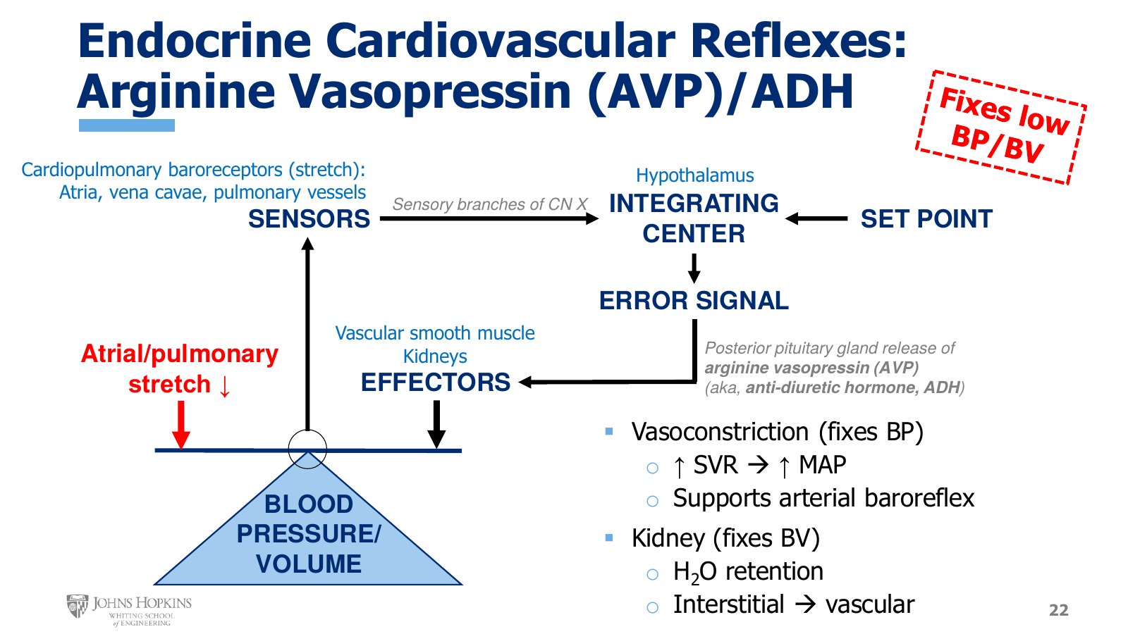
Slide 22. Vasopressin on the negative feedback schematic. Falling atrial and pulmonary stretch, the hypothalamus as integrating center, posterior pituitary release, and effects on vascular smooth muscle and the kidneys. The stamp reads Fixes low BP/BV.
The first endocrine reflex loop involves the hormone arginine vasopressin, also known as antidiuretic hormone, or ADH. A note at the top of each of these slides says whether the hormone addresses low or high pressure and volume. Vasopressin corrects blood pressure, BP, and blood volume, BV, when they are too low.
The negative feedback schematic has blood pressure and blood volume as the regulated variables. Those are separate and distinct variables in the body, so how can they roll into one? The body is actually paying attention to stretch registered in the cardiopulmonary baroreceptors in the walls of the atria, vena cava, and pulmonary vessels. That tells us vasopressin release is truly sensitive to decreases in blood volume, which it is.
For vasopressin, the decrease in stretch at the cardiopulmonary baroreceptors is transmitted through branches of the vagus nerve, cranial nerve X, up to the hypothalamus in the brain. The hypothalamus stimulates the release of vasopressin from the posterior pituitary gland, which sits directly underneath it. Vasopressin enters the bloodstream and acts as the messenger carrying instructions on how to fix the problem, acting on its receptors on vascular smooth muscle and in the kidneys.
Vascular smooth muscle. Vasopressin stimulates a powerful vasoconstriction, which is where it gets the name vasopressin. The vasoconstriction increases systemic vascular resistance, helping correct any decrease in blood pressure that accompanied the decrease in blood volume. This is a bit of a band-aid solution, addressing symptoms rather than causes. But if low blood pressure occurs because of low blood volume, it is absolutely essential: a great hormone companion to the arterial baroreflex, which has undoubtedly already been activated.
Kidneys. Here vasopressin helps fix the underlying root problem of low blood volume. The kidneys determine how much fluid we lose in the urine, and vasopressin tells them to retain water. Water is a valuable molecule right now, not a waste product to be eliminated in the urine. The kidney initially puts retained water into the interstitial space, which then very quickly equilibrates into the vascular space, increasing blood volume.
The renin-angiotensin system: the big picture
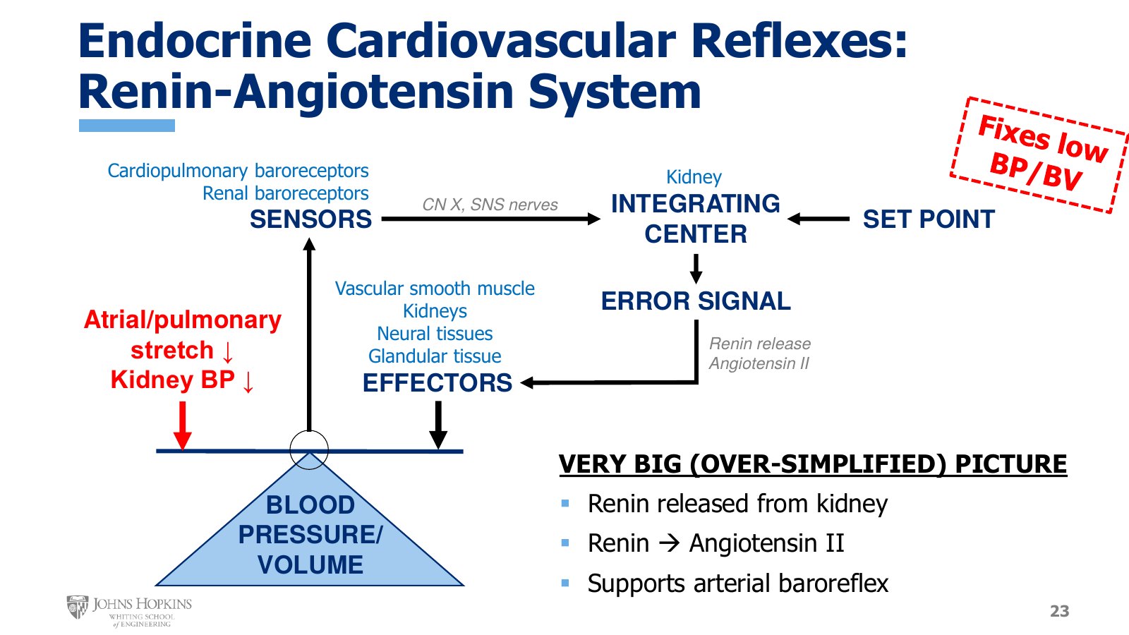
Slide 23. The renin-angiotensin system as a feedback loop, labeled a very big, over-simplified picture. Cardiopulmonary and renal baroreceptors feed the kidney through CN X and sympathetic nerves.
A second major endocrine element of the atrial stretch reflex that addresses low blood volume and blood pressure is the renin-angiotensin system. It is engaged when cardiopulmonary stretch decreases, sensed by the cardiopulmonary baroreceptors, and when blood pressures in the kidneys decrease, sensed by specialized renal baroreceptors. All of this stretch and pressure information converges on specialized cells in the kidneys responsible for releasing renin, the first step in the system.
It is easy to see how information about low blood pressure at the kidney simply sticks around in the kidney. Information from the cardiopulmonary baroreceptors still relies on cranial nerve X to get from the atria to the cardiovascular centers in the medulla. The medulla then sends sympathetic nerve signals to the kidney saying we need to release renin now. And renal blood pressure, let’s face it, is probably low because of low blood pressure upstream in the major systemic arteries.
Once renin is released in response to signals about low blood volume and low renal blood pressure, it unleashes a cascade of events. In a very simplified overview, renin stimulates the formation of angiotensin II, which targets vascular smooth muscle, the kidneys, the nervous system, and endocrine glands to bring about physiological changes that support the arterial baroreflex in restoring blood pressure and blood volume to normal. That would not be exactly wrong, but it is oversimplified enough to be misleading.
The cascade in more detail
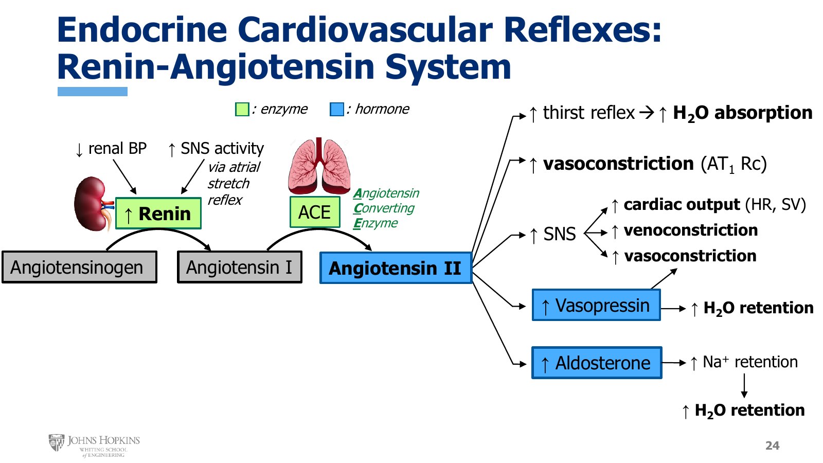
Slide 24. Renin and ACE in green as enzymes, angiotensin II, vasopressin, and aldosterone in blue as hormones. The five branches from angiotensin II end in more water absorption, vasoconstriction, sympathetic effects, and water retention.
The kidney is the source of renin, and the two major stimuli for its release are a drop in renal blood pressure and an increase in sympathetic stimulation of the kidney through the atrial stretch reflex.
A protein called angiotensinogen floats around in the blood plasma all the time. It is inactive and does not do a thing.
When it encounters renin in the blood, renin converts angiotensinogen into angiotensin I. Angiotensin I also does nothing as it hangs out in the blood, but it is an intermediate product, one step closer to activating something.
As blood circulates through the pulmonary circulation, angiotensin I contacts an enzyme found in particularly high concentrations lining the pulmonary blood vessels: ACE, which stands for angiotensin converting enzyme.
ACE converts angiotensin I into the hormone angiotensin II, which is where things really take off.
Angiotensin II is responsible for a whole host of responses, all of which increase either blood pressure or blood volume.
Thirst. It stimulates the thirst reflex in the brain, making us ingest more fluid and expanding blood volume through water absorption in the stomach and intestines.
Vasoconstriction. It binds its AT1 receptors on vascular smooth muscle, stimulating vasoconstriction throughout the systemic circulation, which increases systemic vascular resistance and so blood pressure.
Sympathetic activity. It stimulates sympathetic nervous system activity, which, as we know by this point, increases blood pressure by increasing venoconstriction, vasoconstriction, and cardiac output.
Two more hormones. It stimulates further increases in blood volume and blood pressure by stimulating release of arginine vasopressin and a new hormone, aldosterone.
Vasopressin, as we just saw, increases blood pressure by inducing vasoconstriction and increases blood volume by increasing water retention at the kidney. Aldosterone works similarly, but a little differently. It stimulates retention of sodium ions at the kidney. There is a saying in physiology: where salt, or sodium, goes, water follows. Retaining sodium changes the osmotic balance in the body, making body fluids more concentrated, which promotes secondary osmotic water retention. In both cases, water retained at the kidney quickly equilibrates into the vascular space, expanding blood volume.
Atrial natriuretic peptide
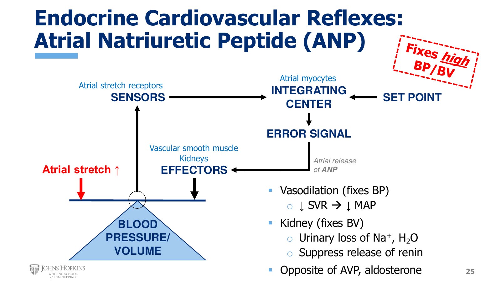
Slide 25. ANP on the negative feedback schematic. Rising atrial stretch, the atrial myocytes as integrating center, and vasodilation, urinary Na⁺ and water loss, and renin suppression as outputs. The stamp reads Fixes high BP/BV.
The last endocrine reflex kicks in when blood pressure and blood volume are too high. The neural Bainbridge reflex is the major way the heart copes with a volume overload. Atrial natriuretic peptide, or ANP, helps the entire body cope with and fix a volume overload.
Blood pressure and blood volume are still the regulated variables. The stimulus is an increase in atrial stretch, sensed by atrial stretch receptors, which stimulate the atrial myocytes to release their stores of ANP. ANP circulates in the bloodstream and binds its receptors on vascular smooth muscle and in the kidneys.
Vascular smooth muscle. ANP stimulates a powerful vasodilation, which decreases systemic vascular resistance and so blood pressure.
Kidney. ANP has a couple of effects here.
It stimulates a urinary loss of sodium ions, called natriuresis. Since where sodium goes water follows, water moves osmotically out of the body and into the urine as well. The result is an overall loss of body water, including water from the vascular space, decreasing blood volume.
It suppresses the release of renin, which keeps the renin-angiotensin system from opposing ANP’s efforts to decrease blood pressure and blood volume.
The endocrine arm of the atrial stretch reflex
Hormone | Fixes | Source | Vessels | Kidney |
|---|---|---|---|---|
Arginine vasopressin (ADH) | Low BP and BV | Posterior pituitary, on hypothalamic signal | Powerful vasoconstriction | Water retention |
Angiotensin II | Low BP and BV | Renin from the kidney, then ACE in the lungs | Vasoconstriction through AT1 | Through vasopressin and aldosterone |
Aldosterone | Low BP and BV | Released on angiotensin II stimulation | None named | Na+ retention, water follows |
ANP | High BP and BV | Atrial myocytes | Powerful vasodilation | Na+ and water loss, renin suppressed |