2A · Introduction to Excitable Membranes
Module 1 established that membranes, by virtue of their phospholipid composition, do a great job of separating fluid compartments and their hydrophilic solutes, maintaining the distinct solution characteristics of the extracellular and intracellular environments. Sodium is concentrated outside the cell, potassium inside. Chloride is more abundant outside, proteins heavily concentrated inside.
What that earlier discussion did not consider, and what matters now, is that those solutes also carry an electrical charge. Some positive, others negative. Wherever a given ion sits at higher concentration, it also exerts a stronger influence over the collective electrical environment of that compartment.
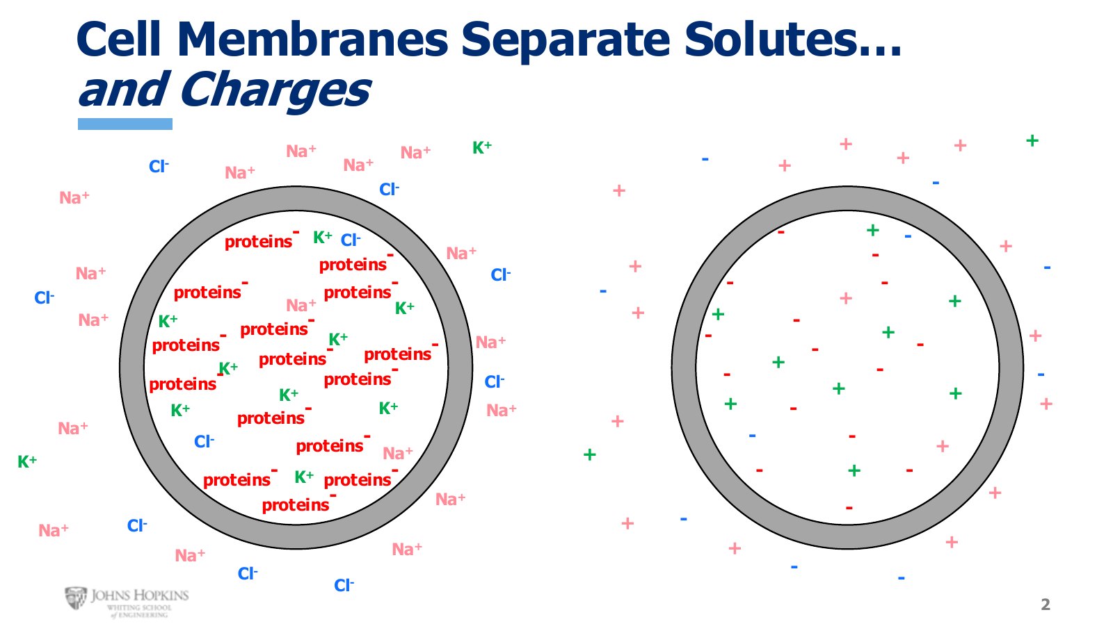
Slide 2. The same solute map from Module 1, then stripped down to charges alone, then to net charge per compartment.
Strip away the ion identities and leave only the charges they carry, and the two compartments turn out to possess distinct electrical characteristics too. Reduce it further to overall net charge and the result holds in pretty much every cell in the body: an electrical gradient is present. Negative charges predominate inside, positive charges predominate outside.
Quantifying the charge difference
By how much? Place very small electrodes in the intracellular and extracellular fluids, with the extracellular electrode always functioning as the ground reference, and a volt meter reports exactly how negative the inside of the cell is compared to the outside.
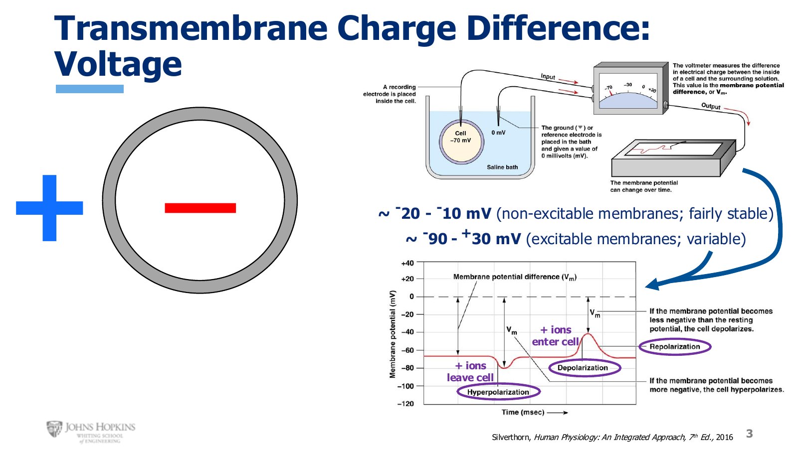
Slide 3. Transmembrane voltage. Non-excitable membranes sit in a narrow band; excitable membranes range across 120 mV.
In most cells in the body that charge difference, expressed in millivolts, is fairly small. The inside sits around 10 to 20 mV more negative than the outside, and that millivoltage is stable and mostly unchanging.
Excitable membranes are different. Their transmembrane charge difference is highly variable and is designed to change, anywhere from −90 mV, meaning the inside is very negative compared to the outside, all the way up to +30 mV on occasion, meaning the inside reverses its polarity and temporarily finds itself more positive than its extracellular environment.
Reading the waveform
Because excitable membranes are built to experience large fluctuations in transmembrane voltage, the object of study becomes the waveform: how voltage changes over time. On a tracing taken from a neuron membrane, the baseline unstimulated value is −70 mV.
If ion channels open that let positive ions leave the cell, taking their positive charges with them, the residual environment inside becomes more negative and the waveform deflects downward. This downward deflection below baseline is hyperpolarization.
If ion channels open that let positive ions enter the cell, the inside becomes less negative, more positive, and the waveform deflects upward. This upward deflection above baseline is depolarization.
When those deflections decay and return to baseline resting values, that is repolarization.
The definition
Excitable membranes are cell membranes designed to experience fluctuations in transmembrane voltage and to capitalize on those fluctuations to do cellular work. They contain a much wider variety of ion channels, so they can precisely control the flow of ions across the membrane. Ion flow changes transmembrane voltage, and that gives a biological electrical signal, which acts as a form of cellular communication.
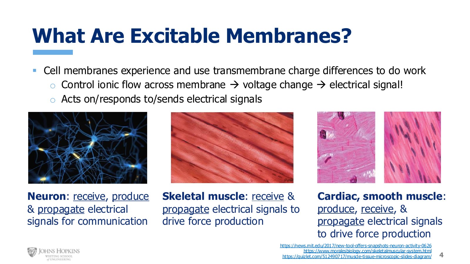
Slide 4. Three excitable tissue types and what each one does with electrical signals. Not every tissue does all three.
What each excitable tissue does with an electrical signal
Tissue | Receive | Produce | Propagate |
|---|---|---|---|
Neuron | yes | yes | yes |
Skeletal muscle | yes | — | yes |
Cardiac and smooth muscle | yes | yes | yes |
Neurons receive electrical signals from other neurons, produce their own, and propagate them to the next cell in the chain of communication. Skeletal muscle cells receive signals from neurons and then propagate them along their own membranes, making sure the entire membrane, which can be quite large in these cells, experiences the change in voltage. That is what drives contraction and force production. Cardiac and smooth muscle produce their own signals, receive signals from other cells, and propagate them onward, driving contraction in the walls of many organs.