2F · Action Potentials
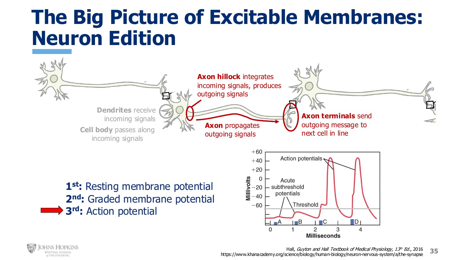
Slide 35. The three neuron states a final time. This lecture is the third: the action potential.
The cell has reached threshold at the axon hillock and commits to sending an outgoing message. This lecture falls into three parts: how action potentials are produced, how they are propagated down the axon, and how they are transmitted to the next cell.
Slide 36. The outline for the lecture: production, propagation, transmission.
The waveform
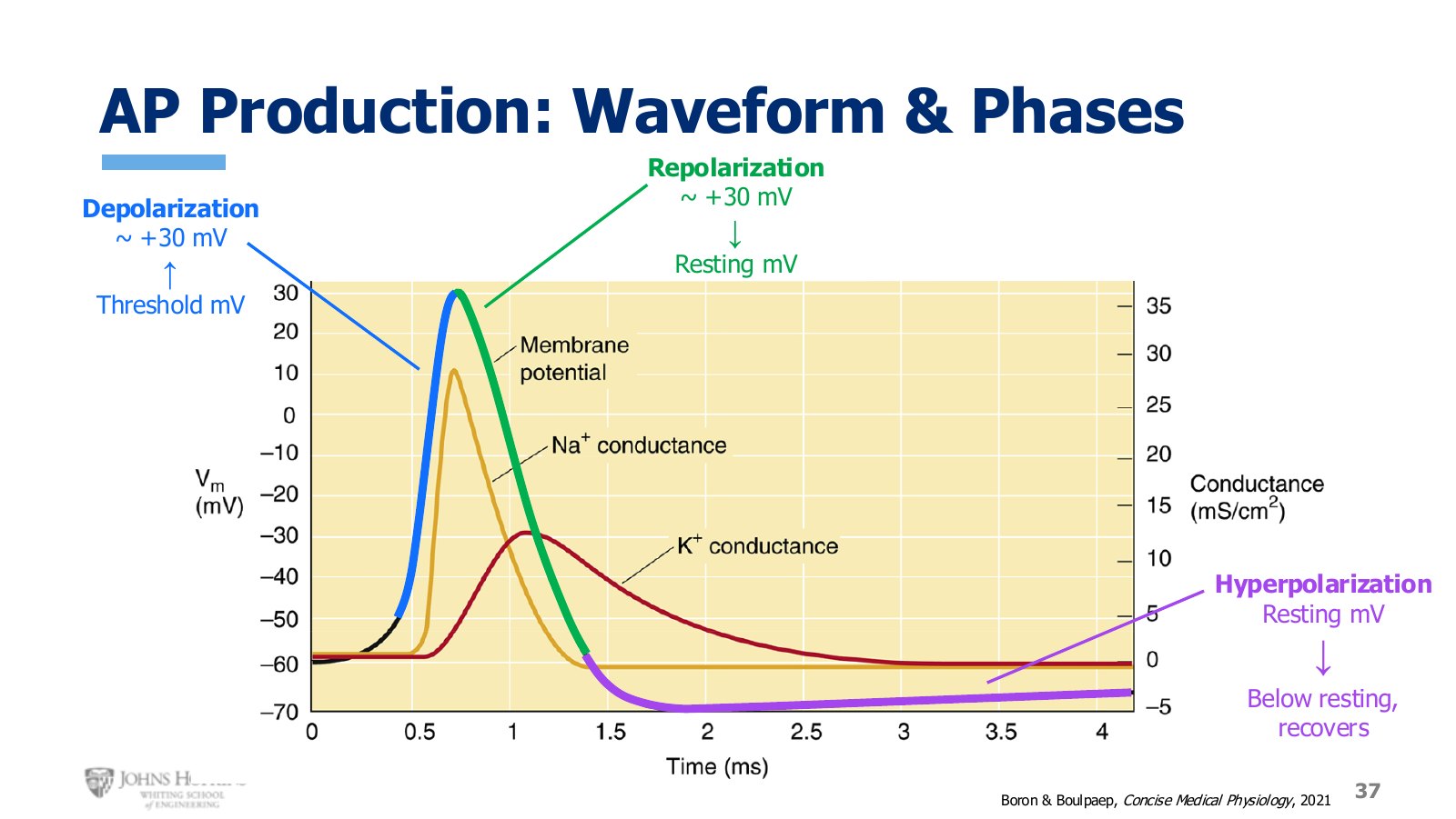
Slide 37. The action potential waveform with its three named phases. Note the one millisecond timescale.
A brief resting period, then a gentle upslope that brings the membrane to threshold, then an explosive upward trajectory followed by an equally sudden return down.
Depolarizing phase: the entire upward slope, up to about +30 mV.
Repolarizing phase: from +30 mV back down to resting.
Hyperpolarization: the membrane goes more negative than resting potential for a little while, then gradually recovers to its original resting membrane potential.
The timescale matters. It takes one millisecond from reaching threshold to the end of repolarization. This is an incredibly fast event in the neuron membrane.
Ion channel events
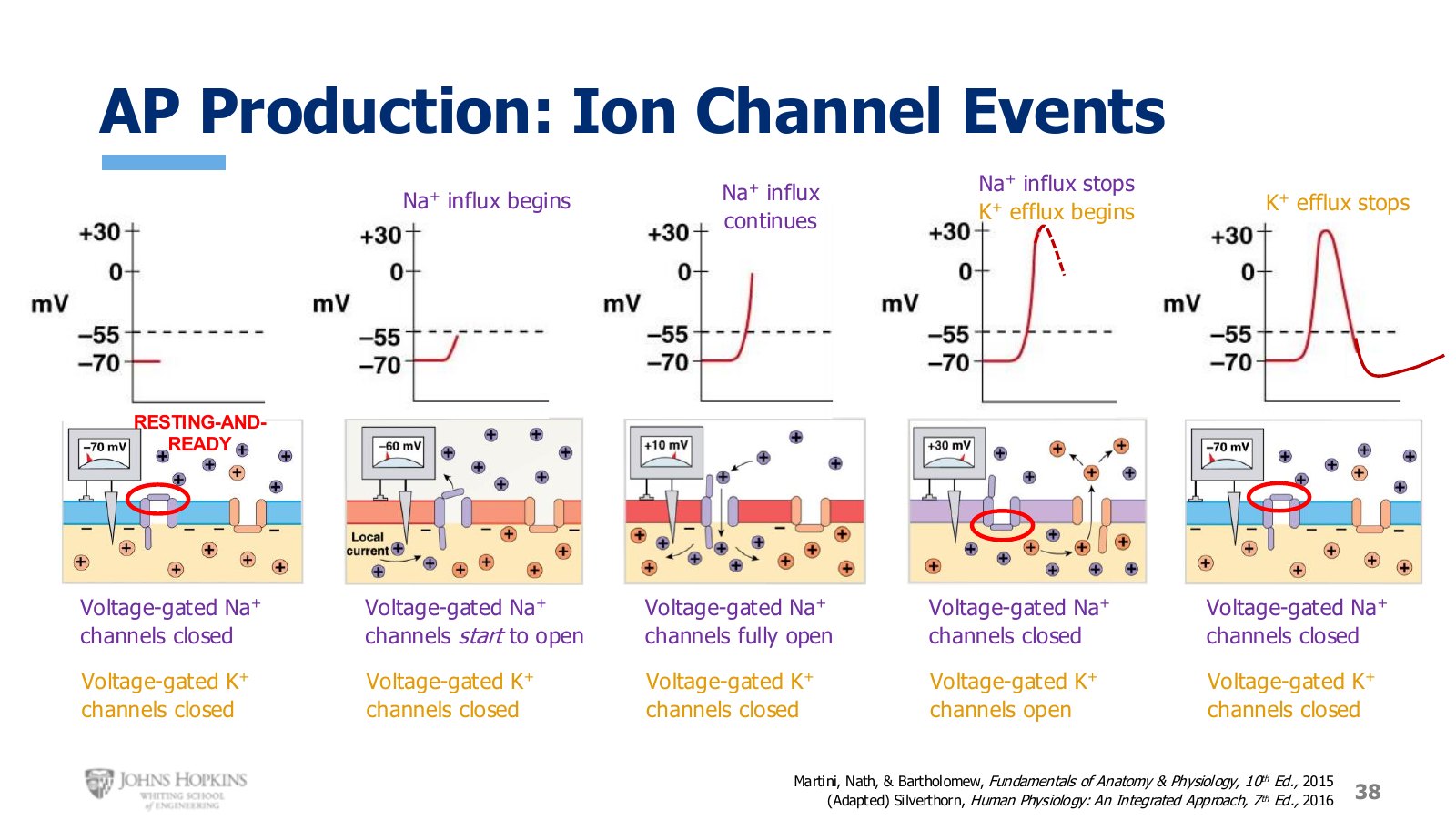
Slide 38. Five stages, with the state of every voltage-gated sodium and potassium channel at each one.
The channels that matter here are voltage-gated. Module 2 has now seen three channel types in sequence: ungated leak channels that establish resting potential, ligand-gated channels that produce graded potentials, and now voltage-gated channels for action potentials. As the name implies, voltage-gated channels require a change in membrane voltage to open. They require depolarization.
Note the structure of the sodium channel. It has an activation gate on the extracellular side and an inactivation gate on the intracellular side. At rest the activation gate is closed and the inactivation gate is open. In this orientation the channel is in its resting and ready conformation: closed, but poised to open easily when stimulated.
At rest. Membrane potential is stable, so both channel types are closed. Sodium channels sit resting and ready.
Threshold. Sodium influx from adjacent upstream depolarizations, such as a graded potential finally reaching the axon hillock, makes membrane potential just positive enough to reach threshold, which nudges voltage-gated sodium channels open. Depolarization begins. Sodium channels respond instantaneously; potassium channels are sluggish and take much longer to open, so they are still closed. That bit is important.
Depolarization. More and more voltage-gated sodium channels open, so sodium influx is strong. Potassium channels remain closed.
Peak, about +30 mV. The rapidly responding sodium channels start to close while the sluggish potassium channels finally start to open. Sodium influx ends, potassium efflux begins. Potassium already tends to leave at rest, and that tendency is only strengthened at +30 mV, where the electrical gradient now reinforces the concentration gradient. Potassium really wants to leave the cell now, and as it does, taking its positive charges with it, repolarization is underway.
Repolarization and hyperpolarization. Potassium efflux finishes repolarization and carries the membrane past resting into hyperpolarization, again because potassium channels are sluggish, this time slow to close. Then potassium channels close, sodium channels finish resetting, and the membrane is ready for the next round.
Refractory periods
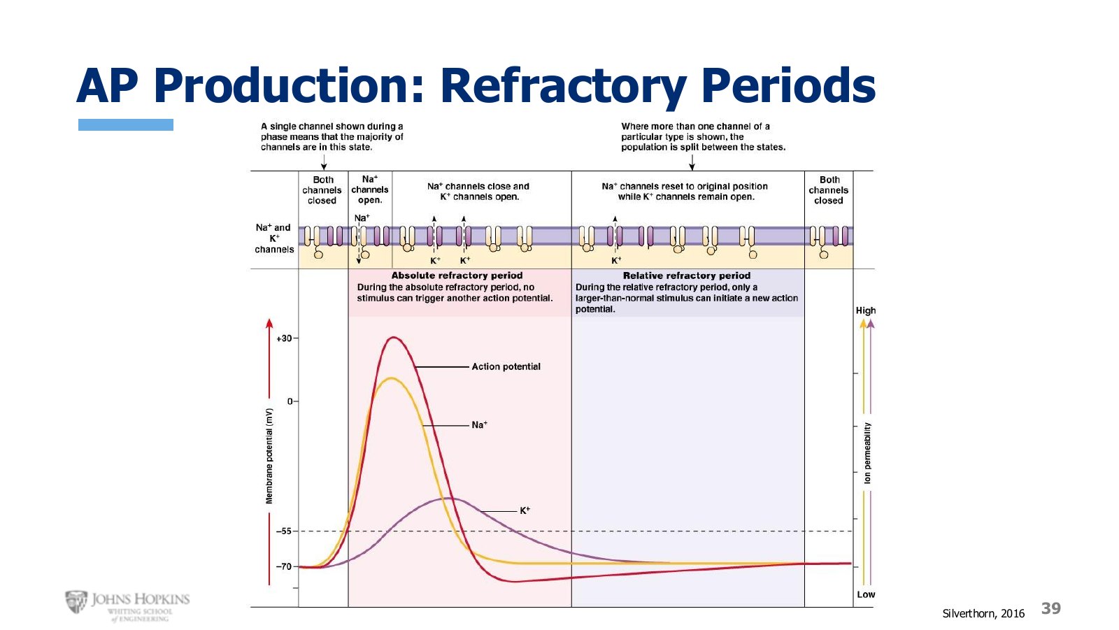
Slide 39. Sodium and potassium permeability against the waveform, with the two refractory periods marked below.
Both the permeability waveforms on this figure and the conductance waveforms on the earlier one correspond to the number of ion channels open at a given time. Lots of sodium channels open almost instantly when depolarization starts, so sodium permeability closely tracks the upward slope, peaks when membrane potential peaks, and declines as sodium channels close through repolarization. Potassium channels are much slower to respond, so their permeability tracing has a delayed onset and a much more gradual upslope and downslope.
The new information is the timescale of the sodium channel conformations. As long as sodium channels are either open, or closed with the inactivation gate closed and the activation gate open, they cannot open again. If the neuron were stimulated again it could not respond, because the sodium channels cannot open a second time. This is the absolute refractory period, so named because there is absolutely no chance another stimulus could lead to another action potential.
Once sodium channels start to reset back to resting and ready, they do it one by one rather than all at once, so the neuron gradually regains the ability to be stimulated. This is the relative refractory period. The neuron is still hyporesponsive and does not really want to respond yet, but it could, if the stimulus is stronger than normal. Threshold may effectively sit at −50 or even −45 mV for this period, until all channels are reset and ready again.
Propagation: electrotonic conduction
Without refreshing the action potential periodically, the signal decays and dies out as it travels farther from the axon hillock. Propagation is the process of refreshing and reiterating new action potentials along the length of the axon so the signal stays strong across the full distance from axon hillock to axon terminals, which can be up to a meter.
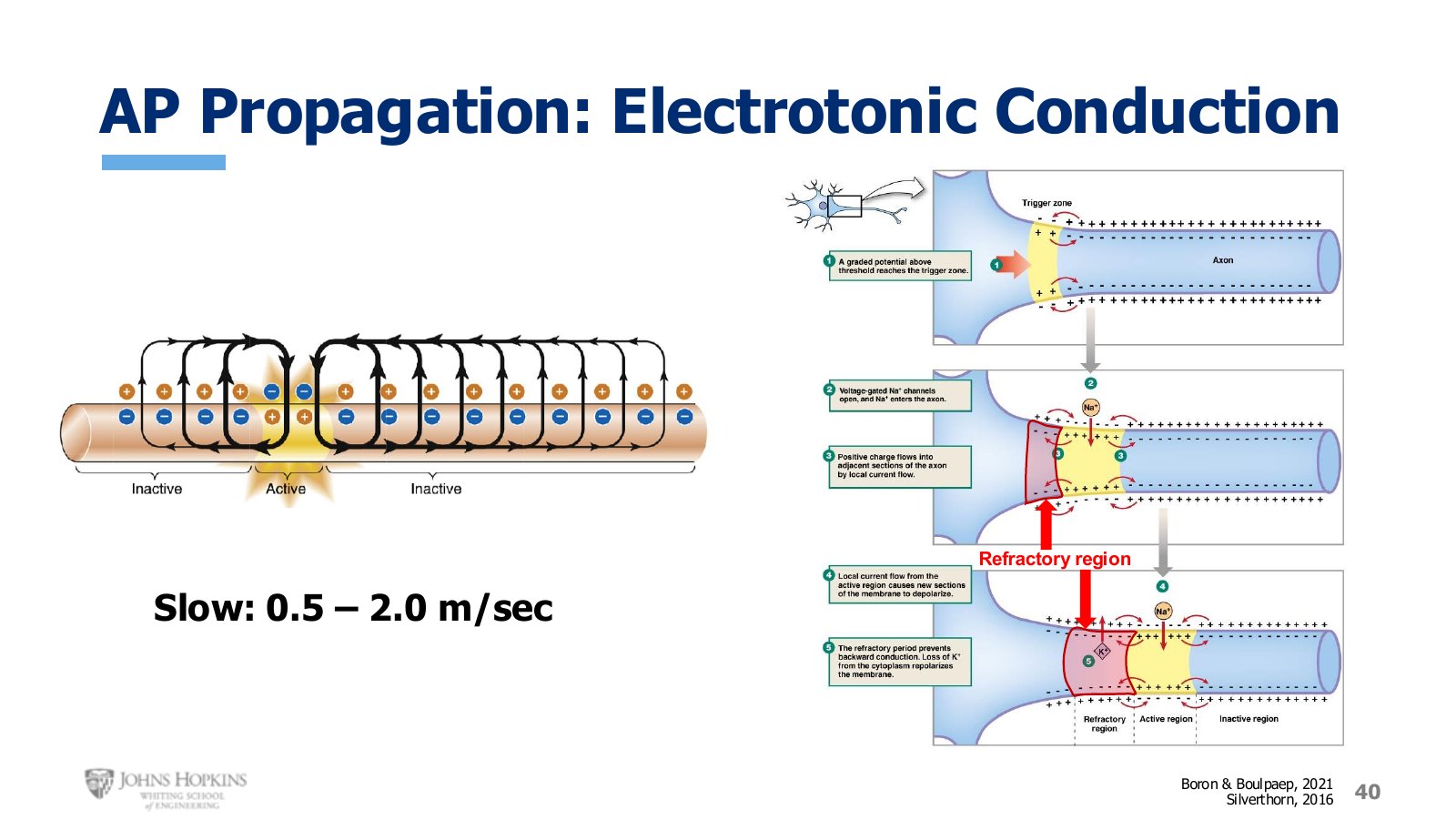
Slide 40. Electrotonic conduction. The refractory region behind the signal is what makes it travel one way.
Electrotonic conduction, which some books call continuous conduction, works a bit like toppling a string of dominos. Tip the first one and it tips the second, which tips the third.
Once the first action potential is triggered at the axon hillock, the huge influx of sodium associated with that localized depolarization diffuses over to the next adjacent portion of the axon, bringing its local membrane potential to threshold and triggering an action potential there. When that second region depolarizes, some sodium diffuses upstream and some downstream. The upstream portion cannot be stimulated again because it is currently in its refractory period, so functionally the signal can only travel downstream. The process repeats many times until the signal reaches the axon terminals.
As you would expect, this is time-consuming. Electrotonic propagation travels at anywhere from 0.5 to 2 m/s.
Propagation: saltatory conduction
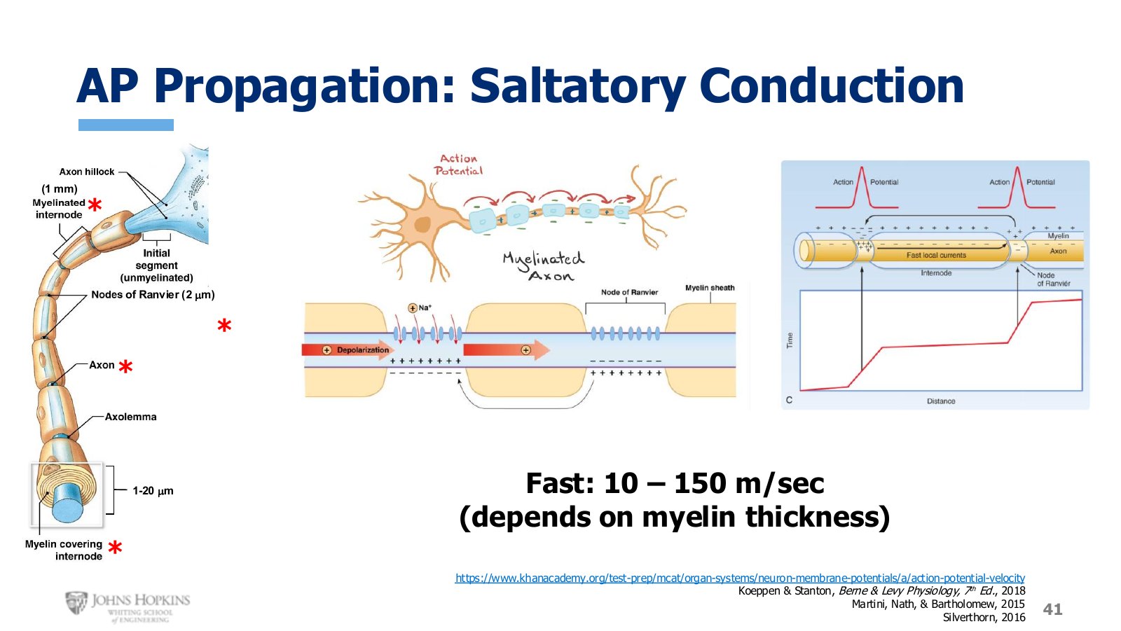
Slide 41. Myelin sheaths and the nodes of Ranvier between them. The signal refreshes only at the nodes.
The second form of propagation adds a structure and a cell type. Myelination is produced when a very thin flat cell, a Schwann cell in the peripheral nervous system or an oligodendrocyte in the central nervous system, wraps itself around the axon many times, like rolling up a flattened tube of toothpaste.
It wraps 20 to 300 layers around an axon of 1 to 20 micron diameter, becoming a form of electrical insulation and increasing transmembrane resistance. A single myelin sheath runs about one millimeter, followed by a small naked piece of axon called a node of Ranvier, only about two micrometers long. Then another sheath, another node, and so on.
Covering most of the axon with insulation and exposing only small periodic sections means the action potential does not have to travel laboriously down every section. It jumps from node to node, leap-frogging its way down. Saltatory is derived from the French word for jump.
The jump works because voltage-gated ion channels sit only in the membrane at the nodes, so nodes are the only places an action potential can be produced. The myelin means the strength of the depolarizing signal does not dissipate between nodes, because insulation makes ion flow across the membrane impossible. Transit is very fast across myelinated sections, since the action potential only needs to refresh itself periodically. You can see the time cost of crossing each naked node, then the signal covers a much longer distance in a fraction of the time.
Myelinated axons conduct at 10 to 150 m/s, with conduction velocity proportional to the thickness of the myelin wrapping.
What sets conduction velocity
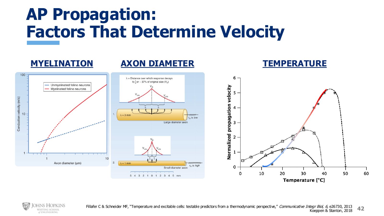
Slide 42. Three variables: myelination, axon diameter, and temperature.
Myelination. Myelinated conduction velocity is simply much higher than unmyelinated.
Axon diameter, meaning the underlying axon itself minus any myelin wrapping. Conduction is faster in thicker axons. Longitudinal resistance down the axon is higher in thinner axons and lower in thicker ones, so the signal travels further, and thus faster, before it decays.
Temperature. Higher temperatures lead to faster propagation, particularly within physiological limits. Warming cat neurons from 10°C to 40°C takes propagation velocity from nearly nothing at 10°C, through a reference baseline at 20°C, up to five times that baseline at 40°C. Module 1 established that high temperatures speed up diffusion in general, and that is playing out here in the diffusive flow of ions across the membrane and down the axon.
Transmission: synaptic events
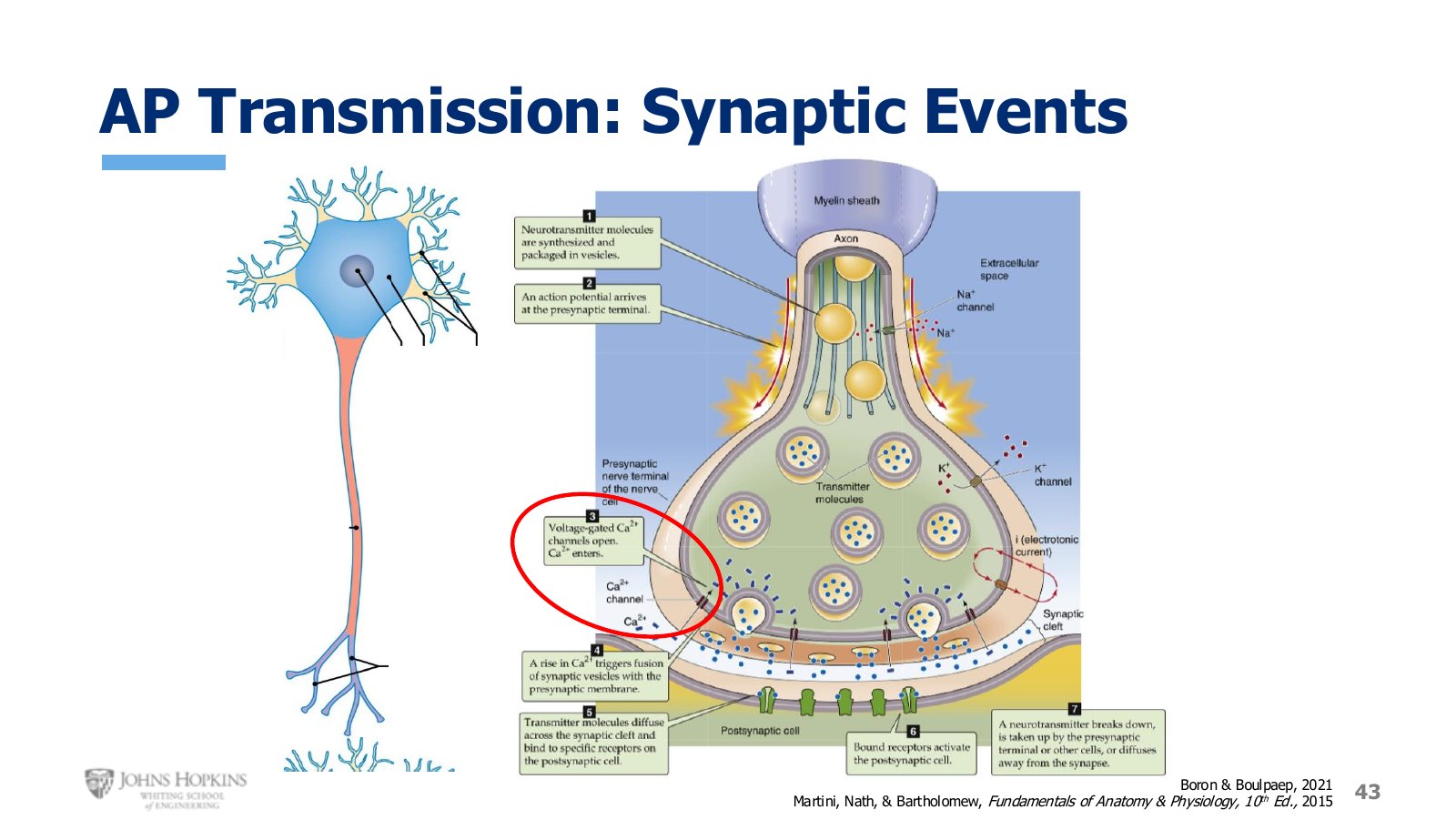
Slide 43. The axon terminal in close-up. Calcium entry is what converts the electrical signal back into a chemical one.
As the action potential travels down the membrane of the axon terminals, the wave of depolarization opens additional voltage-gated channels. Voltage-gated sodium and potassium channels still open, because that is what creates the action potential, but voltage-gated calcium channels are added now. The action potential triggers them to open, and because calcium has a very strong inward electrochemical gradient, it rushes into the end of the axon terminal.
Calcium plays many roles in the body. It drives muscle contraction, helps set where threshold millivoltage sits, enables blood clotting, and acts as a second messenger inside cells telling them what to do. That last role is what it is doing here.
Calcium attaches to packaged vesicles full of neurotransmitters. Binding to the outside of a vesicle is the signal for it to fuse with the membrane and empty its contents into the synaptic cleft, a process called exocytosis. The neurotransmitters diffuse across the cleft and bind receptors on the postsynaptic membrane, delivering the message to the next cell in line.
Transmission: what the receiving cell has
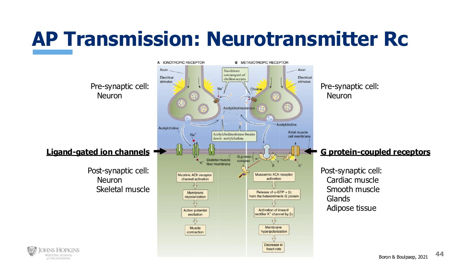
Slide 44. Which receptor the neurotransmitter meets depends entirely on what kind of cell is receiving.
Up to now the postsynaptic cell has been assumed to be another neuron, which simplifies the picture. It could also be a cell in a target tissue: muscle, glands, or fat. The receptor the neurotransmitter binds depends on which.
Postsynaptic receptor by cell type
Postsynaptic cell | Receptor | What happens |
|---|---|---|
Neuron, skeletal muscle | Ligand-gated ion channels | Induces a graded potential, often leading to initiation of an action potential on the membrane. |
Cardiac muscle, smooth muscle, glands, adipose tissue | G protein-coupled receptors | Delivers the message in a very different way, with different cellular outcomes. |
Skeletal, cardiac, and smooth muscle as postsynaptic cells are upcoming topics later in this course.