3A · Skeletal Muscle Anatomy
All muscle tissue exists to generate force to produce movement somewhere in the body. Skeletal muscle, attached to our bones, controls the movement of the body as we interact with the outside world, moving and stabilizing the positions of our limbs, trunk, neck, and head.
Module 2 established that skeletal muscle is innervated by somatic motor neurons. Those neurons are voluntary, meaning the brain can consciously instruct them to fire, so stimulation of skeletal muscle is voluntary too. We can decide to wiggle our toes or arch our backs or smile.
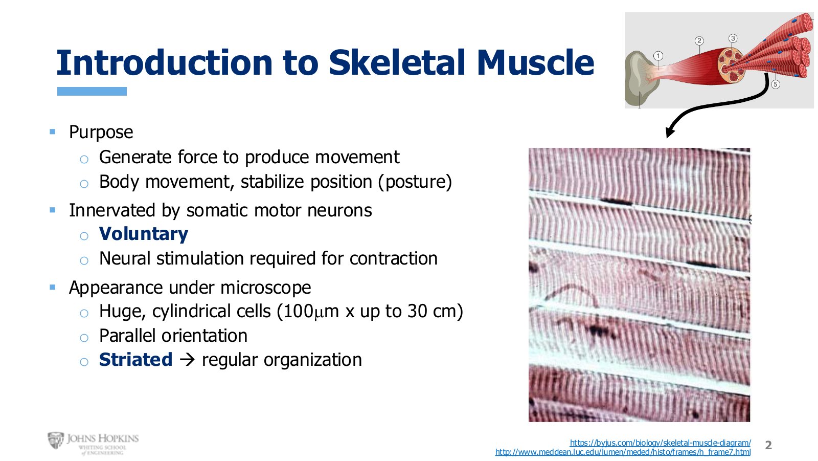
Slide 2. Skeletal muscle under the microscope. Cells run parallel, far longer than they are wide, and visibly striped.
The macroscopic whole muscle attached to a bone is what most of us picture, but it is not where this module spends its time. The focus is inside individual skeletal muscle cells.
Those cells are cylindrical and very large, arranged parallel to one another. On average about 100 microns wide, which is really big for a cell in the body, and reaching up to 30 centimeters long, because they run the entire length of a muscle from tendon to tendon. They are also visibly striped, or striated, and that pattern of light and dark stripes indicates a highly organized intracellular environment.
Structural hierarchy: whole muscle down to cell
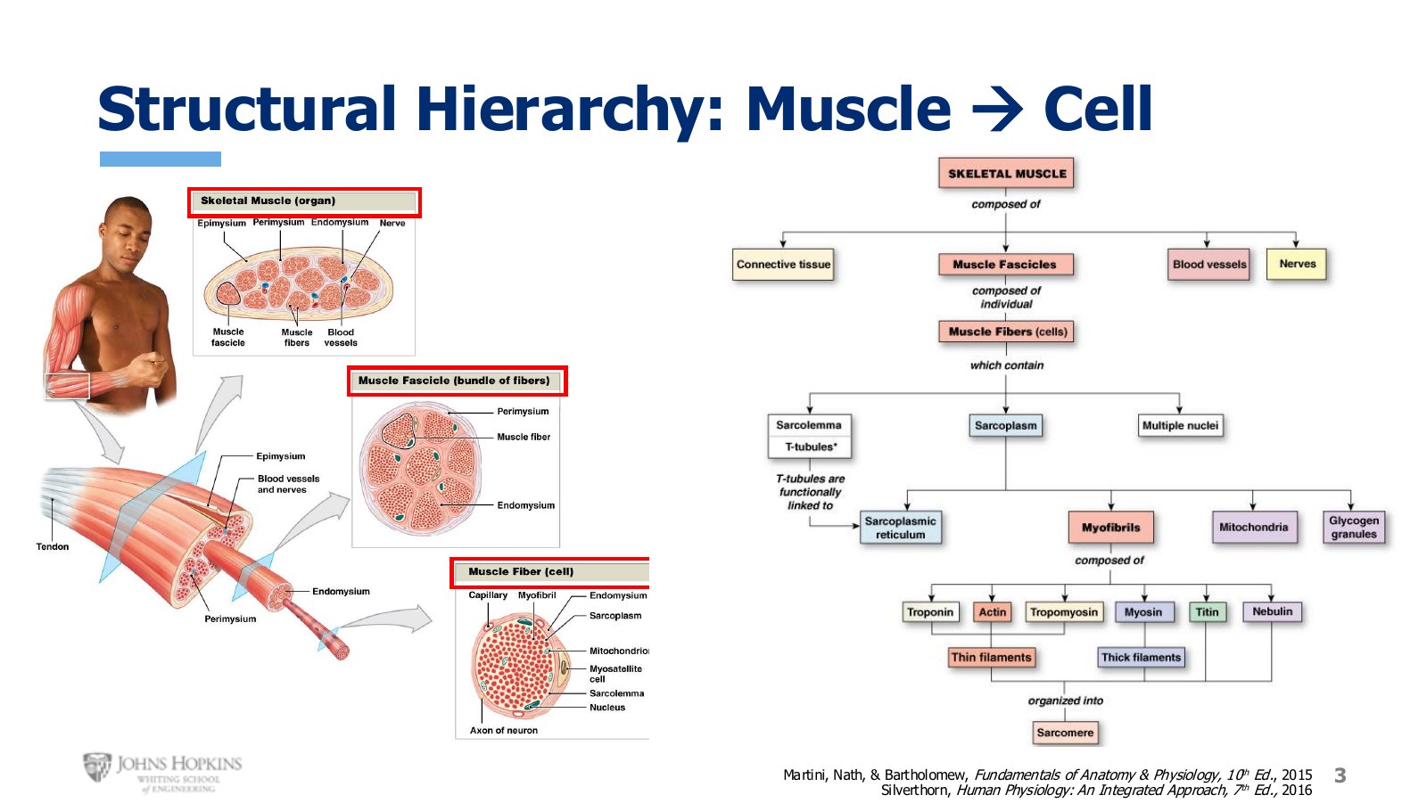
Slide 3. Whole muscle, fascicle, and single fiber, each with its own connective tissue wrapping.
A whole muscle, a biceps brachii or one of the gluteal muscles, is composed of connective tissue, blood vessels, nerves, and bundles of muscle fascicles. The connective tissue shrink wrap holding the whole muscle together is the epimysium.
Each fascicle is a bundle of several muscle fibers, also called muscle cells, shrink wrapped together inside a connective tissue wrapping called the perimysium. Nerves usually run right next to blood vessels.
And each muscle cell is itself made of smaller bundled structures called myofibrils.
Inside the cell
Much of the vocabulary from here starts with sarco- or myo-. Sarco is the Greek prefix meaning muscle; myo is the Latin prefix meaning muscle. These are terms unique to muscle tissue.
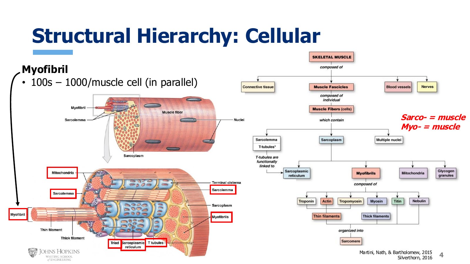
Slide 4. The muscle cell in cross section, with myofibrils packed in parallel and the sarcoplasmic reticulum wrapping each one.
Sarcolemma, the muscle cell membrane, also called a plasmalemma. An excitable membrane.
Several nuclei, because muscle cells are huge.
Sarcoplasm, the muscle-specific name for the intracellular fluid cytoplasm.
Sarcoplasmic reticulum, a muscle-specific version of endoplasmic reticulum. It stores calcium ions.
Lots of mitochondria, which produce ATP for the muscle to use.
Glycogen, intracellular stores of glucose used as fuel for making ATP.
Myofibrils, the contractile machinery.
Zooming in on one portion of a single cell: the sarcolemma sits on the outside, and yellow extensions of it branch into a network of tubes penetrating into the cell. These deep invaginations are transverse tubules, or T tubules, and they carry action potentials deep into the core of the muscle.
The sarcoplasmic reticulum forms a mesh that wraps around each and every myofibril. There are 100 to 1,000 myofibrils inside each muscle fiber, arranged in parallel, independently carrying out the same contractile activity, each running the entire length of the cell.
Inside the myofibril
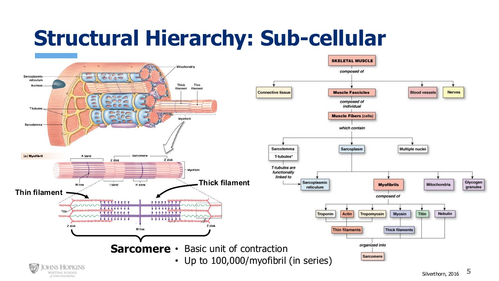
Slide 5. The sarcomere, bounded by Z discs. Thick and thin filaments interdigitate.
Inside each myofibril are specialized cytoskeleton proteins that give skeletal muscle its shape, structural integrity, and contractile function. They are woven, braided, and bonded together into visible thick filaments and thin filaments.
The filaments are anchored in different places so they are positioned to slide past each other. Thin filaments anchor to a network of structural proteins called Z discs, or Z lines. Thick filaments anchor to a different network called M lines.
The smallest level of organization encompassing all of it is the sarcomere. Sarcomeres are arranged end to end in series down a single myofibril, up to 100,000 in length, and physiologists consider the sarcomere the basic unit of contraction. Whatever happens in one sarcomere during contraction and relaxation is happening inside every sarcomere, in every myofibril, in that muscle cell.
Reading a sarcomere
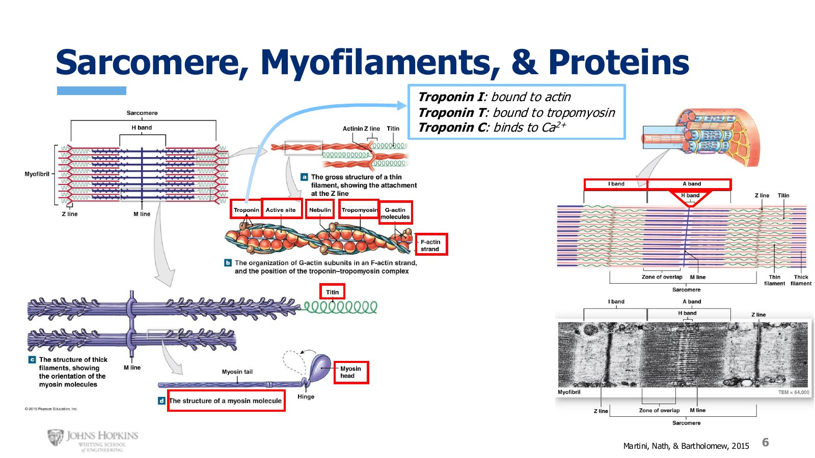
Slide 6. Sarcomere regions and the proteins of each filament, with the three troponin subunits called out.
Z disc, where the thin filaments anchor and project inward toward the center of the sarcomere.
M line, where the thick filaments anchor and project outward toward the sides.
A band, the entire width of the thick filaments, corresponding to the thick dark region under the microscope.
H band, the middle of the A band, where only thick filaments sit with no thin filament overlap.
I band, the region spanning two adjacent sarcomeres where only thin filaments sit, corresponding to the lighter region with the Z disc running down the middle.
The alternation of the lighter I band with the darker A band is what produces the vivid striations that characterize skeletal muscle under the microscope.
The contractile proteins
Thick filaments consist of two proteins bound together. The core is a long protein molecule called titin, which functions as a coiled spring and is responsible for muscle’s passive elastic properties. Around that core sit many myosin molecules, each shaped like a golf club. The long handles, the myosin tails, orient longitudinally along the filament, while the myosin heads stick out radially around the entire circumference.
Thin filaments have a long thin scaffolding protein called nebulin at the core. Everything else orients around it, starting with actin. As individual small spherical molecules it is G-actin; when many G-actin molecules coalesce into a longer strand, like pearls strung into a necklace, it becomes F-actin. Each thin filament has two F-actin strands winding around each other.
Actin and myosin must physically interact to bring about contraction, and they do so at the active site, sometimes called the binding site, where the myosin head grabs onto actin. Most of the time that site is covered by a long rope-like protein called tropomyosin, preventing interaction. That is what produces the quiet resting relaxed state of most muscles.
Moving tropomyosin off the active site is the job of troponin, which is not one molecule but a complex of three subtypes.
The troponin complex
Subunit | Binds to | Role |
|---|---|---|
Troponin I | Actin | Anchors the whole complex to actin. |
Troponin T | Tropomyosin | Controls the positioning of tropomyosin. |
Troponin C | Calcium | Binds Ca2+, the signal to move tropomyosin off the active site. |
Geometry in three dimensions
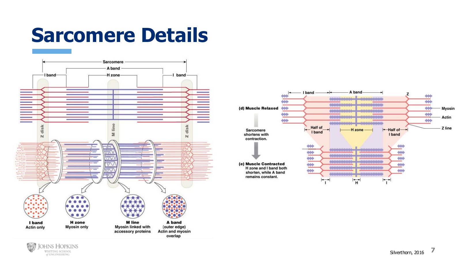
Slide 7. Sarcomere detail, including the cross-sectional packing of filaments.
The filament arrangement is just as precisely orchestrated in cross section as it is longitudinally, with each thick filament surrounded by six thin filaments. That three-dimensional sense helps later when picturing how the filaments slide.
When the muscle is relaxed, each sarcomere is fairly elongated with only a little overlap between thick and thin filaments. During contraction the sarcomere shortens as the thin filaments are drawn toward the center, giving more complete overlap.