3B · Electrical Stimulation at the Neuromuscular Junction
The neuromuscular junction is the specialized synapse between the presynaptic motor neuron and the postsynaptic muscle cell. This lecture covers how a skeletal muscle cell receives the excitatory signal from its upstream neuron.
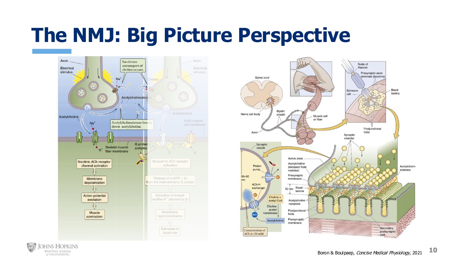
Slide 10. The neuromuscular junction at low magnification, then the motor neuron traced from spinal cord out to the muscle.
Module 2 ended on a figure showing two synapse types. The left side is the broad picture of the neuromuscular junction: an upstream excitatory neuron releases the excitatory neurotransmitter acetylcholine, which binds ligand-gated ion channels, depolarizing the postsynaptic skeletal muscle cell membrane and leading to contraction.
Tracing further upstream, the somatic motor neuron originates in the spinal cord and runs out to the muscle tissue, where the end of the neuron branches to stimulate several skeletal muscle cells at once. At a single synapse, many vesicles of neurotransmitter sit in the axon terminal, waiting to undergo exocytosis and release their acetylcholine into the synaptic cleft.
The sequence
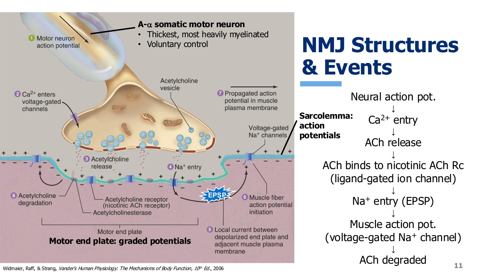
Slide 11. Every step at the junction, from neural action potential down to acetylcholine degradation.
The neuron carrying the message is an A-alpha somatic motor neuron. The A-alpha part tells us this neuron has the thickest and most heavily myelinated axon of any neuron in the body, so the neurons that stimulate skeletal muscle are the fastest neurons we have. They are somatic, so we have voluntary control over them, and therefore over skeletal muscle stimulation.
The neural action potential reaches the axon terminals and opens voltage-gated calcium channels.
Calcium enters the terminal and stimulates release of acetylcholine from its storage vesicles.
Acetylcholine crosses the synaptic cleft and binds acetylcholine receptors on the motor end plate, the specialized region of sarcolemma right at the junction. It contains different ion channels and can only produce graded potentials.
Those receptors are ligand-gated ion channels, also called nicotinic acetylcholine receptors. They open and sodium rushes in.
Sodium influx locally depolarizes a small portion of the motor end plate, producing an excitatory postsynaptic potential.
The EPSP travels to where the motor end plate transitions into regular sarcolemma, which is also where the graded potential is integrated. If it reaches threshold it opens voltage-gated sodium channels and initiates an action potential.
That action potential propagates along the entire sarcolemma in all directions.
Once the neuron stops signalling, residual acetylcholine in the cleft is broken down by the enzyme acetylcholinesterase, preventing ongoing stimulation after the neuron has stopped.
The muscle action potential
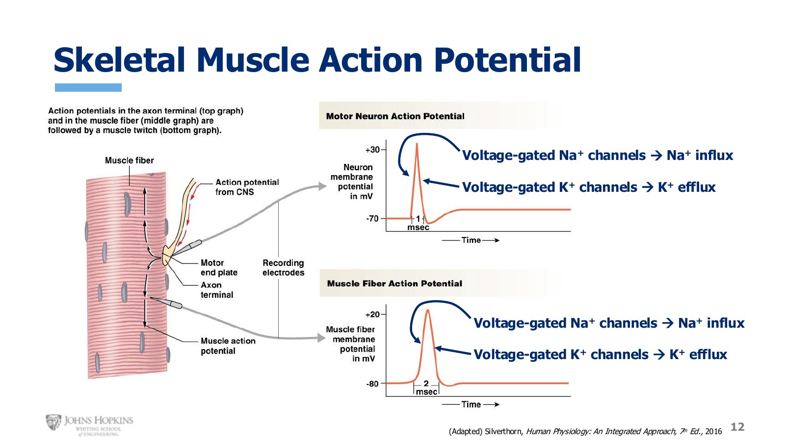
Slide 12. Neural and skeletal muscle action potentials side by side. Same shape, same mechanism, different timescale.
Skeletal muscle action potentials are remarkably similar to neural ones, both in waveform profile and underlying mechanism. The muscle version takes slightly longer, two milliseconds compared to one, but it has the same steep depolarization driven by sodium influx through voltage-gated sodium channels, followed by steep repolarization driven by potassium efflux through voltage-gated potassium channels.
Disruptions at the junction
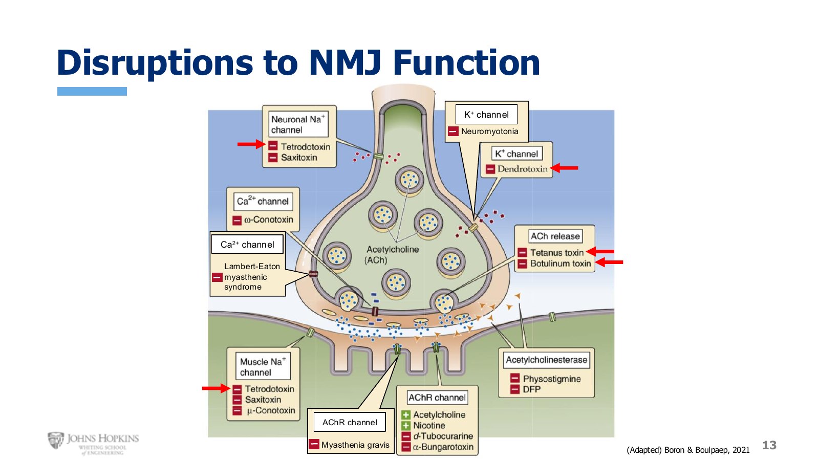
Slide 13. Where toxins and autoimmune conditions attack the junction.
Toxins. Tetrodotoxin from pufferfish is a potent inhibitor of excitable membranes, blocking voltage-gated sodium channels in both neurons and skeletal muscle, leading to instant and lethal paralysis. Dendrotoxin, found in mamba snake venom, inhibits neural voltage-gated potassium channels, which minimizes repolarization and lets depolarization go on far too long. The result is a huge over-release of acetylcholine, relentless depolarization of the sarcolemma, painful cramping and convulsions, then death.
Tetanus toxin, from the bacteria that thrive in deep metallic puncture wounds, and botulinum toxin, which causes botulism food poisoning, both inhibit acetylcholine release, preventing neural stimulation of contraction and leading to death by paralysis of respiratory muscles. In very small targeted doses botulinum toxin is used cosmetically, marketed as Botox, and to treat conditions including migraine headaches and hyperhidrosis.
Autoimmune conditions. These occur when the immune system produces antibodies that attack our own tissues, slowly destroying them over time.
Three autoimmune conditions of the junction
Condition | Target | Consequence |
|---|---|---|
Myasthenia gravis | Acetylcholine-gated ion channels on the motor end plate | Progressive muscle weakness and eventual paralysis. The most common of the three. |
Neuromyotonia | Neuronal voltage-gated potassium channels | Slow destruction of the channels that repolarize the neuron. |
Lambert-Eaton myasthenic syndrome | Voltage-gated calcium channels | Eventually prevents neurotransmitter release. |