3C · Excitation-Contraction Coupling and the Sliding Filament Theory
The presence of calcium ions in the sarcoplasm is essential for contraction to occur. Everything in this lecture runs through that fact.
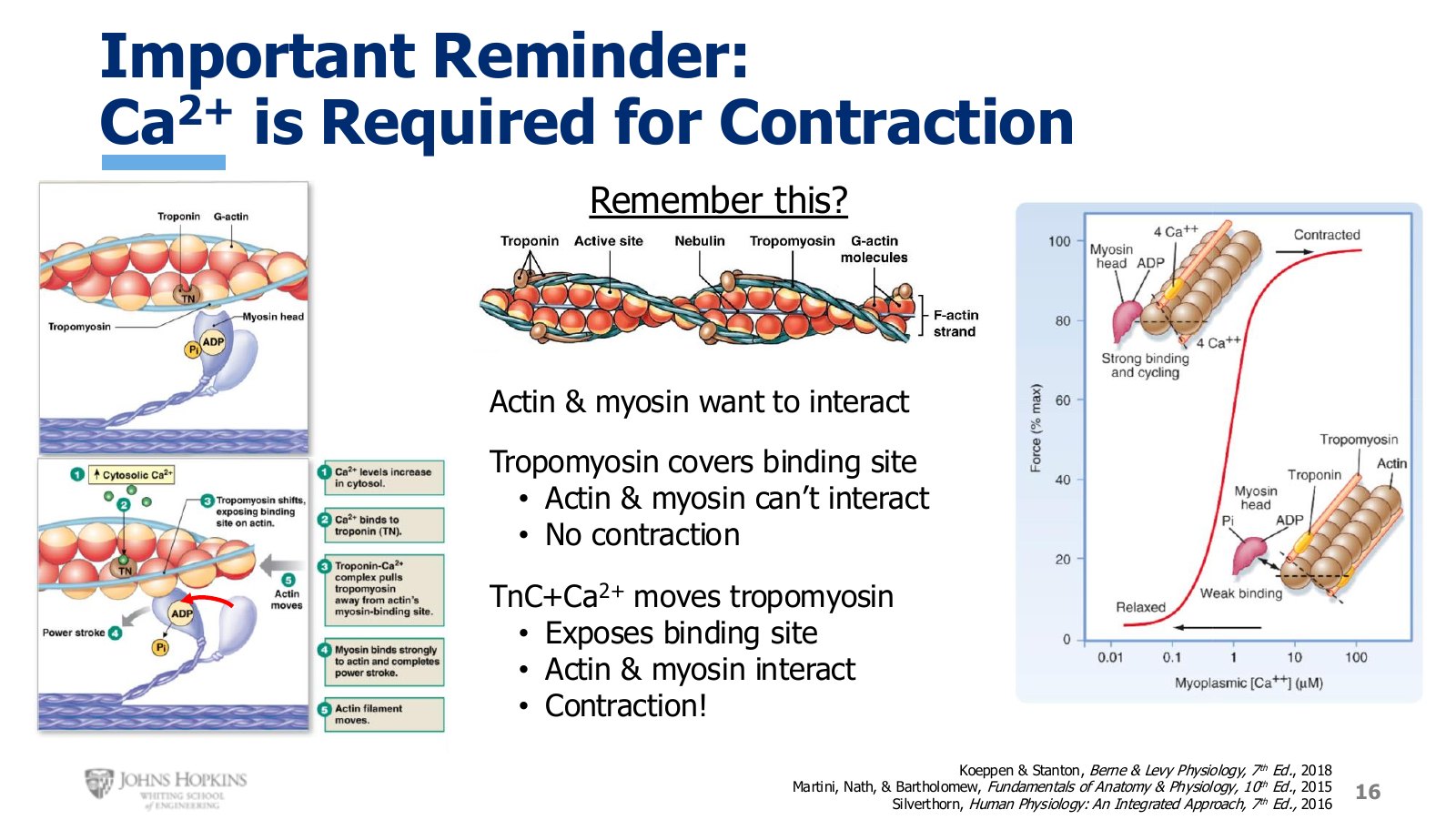
Slide 16. Tropomyosin covers the binding site until calcium binds troponin C and moves it aside.
The thin filament is several proteins bound together: long strands of actin intertwined with tropomyosin and troponin. Actin and myosin have high affinities for each other and really want to bind, with myosin’s head attaching to actin’s binding site. But most of the time tropomyosin covers that site, so myosin cannot grab on, and with no physical point of connection there is no contraction.
When free calcium ions flood into the sarcoplasm, calcium binds troponin C, which makes the troponin complex change shape and pull tropomyosin off the binding site. With the site exposed, actin and myosin bind, and contraction and force generation follow.
Where the calcium comes from
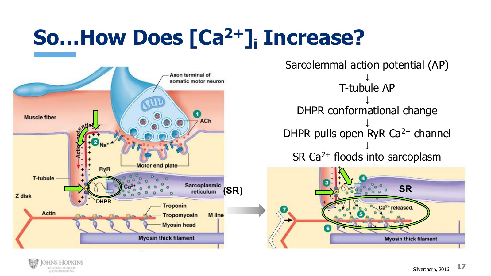
Slide 17. The T tubule action potential deforms the DHPR, which mechanically pulls the ryanodine receptor open.
Pick up with the action potential on the sarcolemma from lecture 3B. It travels along the membrane until it reaches a T tubule. T tubules are simply extensions of the sarcolemma, so the action potential travels down the T tubule membrane.
In the T tubule membrane sit voltage-sensitive receptors called dihydropyridine receptors, or DHPRs. As the wave of depolarization hits them they change conformation. Those DHPRs are physically connected to the ryanodine receptor calcium channels, or RyRs, on the sarcoplasmic reticulum.
This is where the triad’s close physical proximity matters. The conformational change in the DHPR pulls open the calcium channel gate on the sarcoplasmic reticulum, and calcium floods down its concentration gradient out of storage and into the cytoplasm, where it binds troponin C and lets myosin and actin finally bind together.
The sliding filament theory
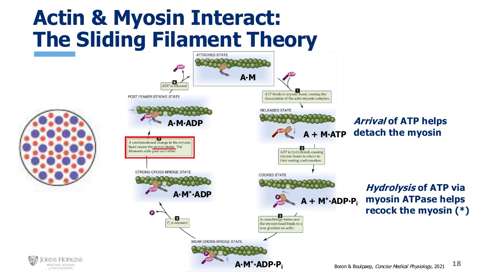
Slide 18. The cross-bridge cycle in shorthand. One ATP molecule does two separate jobs.
The cycle is iterative, so you can enter it anywhere. Start where the myosin head is strongly attached to actin, written A·M. The head has just completed one round of pulling the thin filament toward the center of the sarcomere.
ATP arrives and binds the myosin head, which lets the head detach from actin. Shorthand: A + M·ATP.
ATP is hydrolyzed, and that energy recocks the head into a position full of potential energy, much like a stretched rubber band or compressed spring. Shorthand: A + M*·ADP·Pi, where the asterisk marks the energized head.
The head grabs another G-actin molecule farther up the thin filament than the one it released. This connection is a cross-bridge. Binding is weak at first and gets much stronger once myosin kicks off the inorganic phosphate. Shorthand: A·M*·ADP.
The power stroke. The head undergoes a conformational change and pivots toward the middle of the sarcomere, dragging the thin filament with it and letting the filaments slide past each other. The potential energy is spent, so the asterisk disappears: A·M·ADP.
ADP is kicked off and we are back where we started at A·M.
This cycle runs with every myosin head, in every sarcomere, in every myofibril inside the cell. Given the cross-sectional geometry, a single thick filament can grab up to six thin filaments, and multiple thick filaments can grab a single thin filament. The ongoing power strokes from every cross-bridge in the entire cell are what generate the force we associate with muscle contraction.
Relaxation
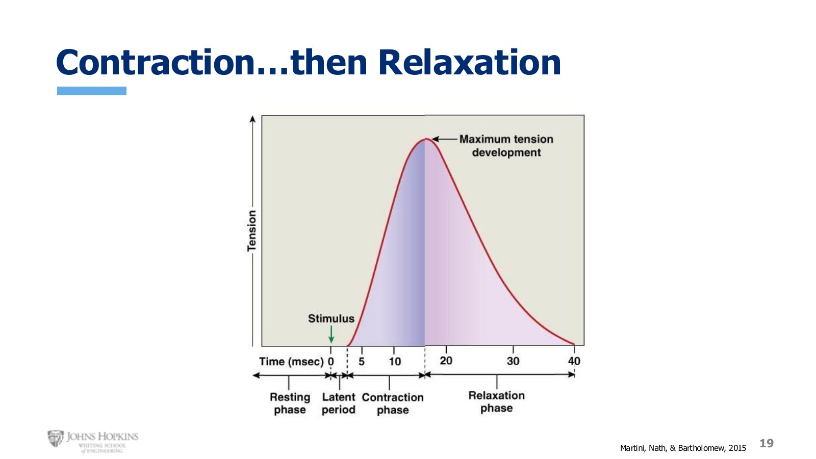
Slide 19. A single twitch is a contraction phase and a relaxation phase. What goes up must come back down.
A twitch contraction waveform plots muscle tension against time: a resting period, the onset of the electrical stimulus, a short latent period while the action potential travels across the sarcolemma and down the T tubules to release calcium, then rising tension as actin and myosin run many iterations of the cycle. But that is only half of it. A twitch consists of both the rising contractile phase and the release of tension during the relaxation phase.
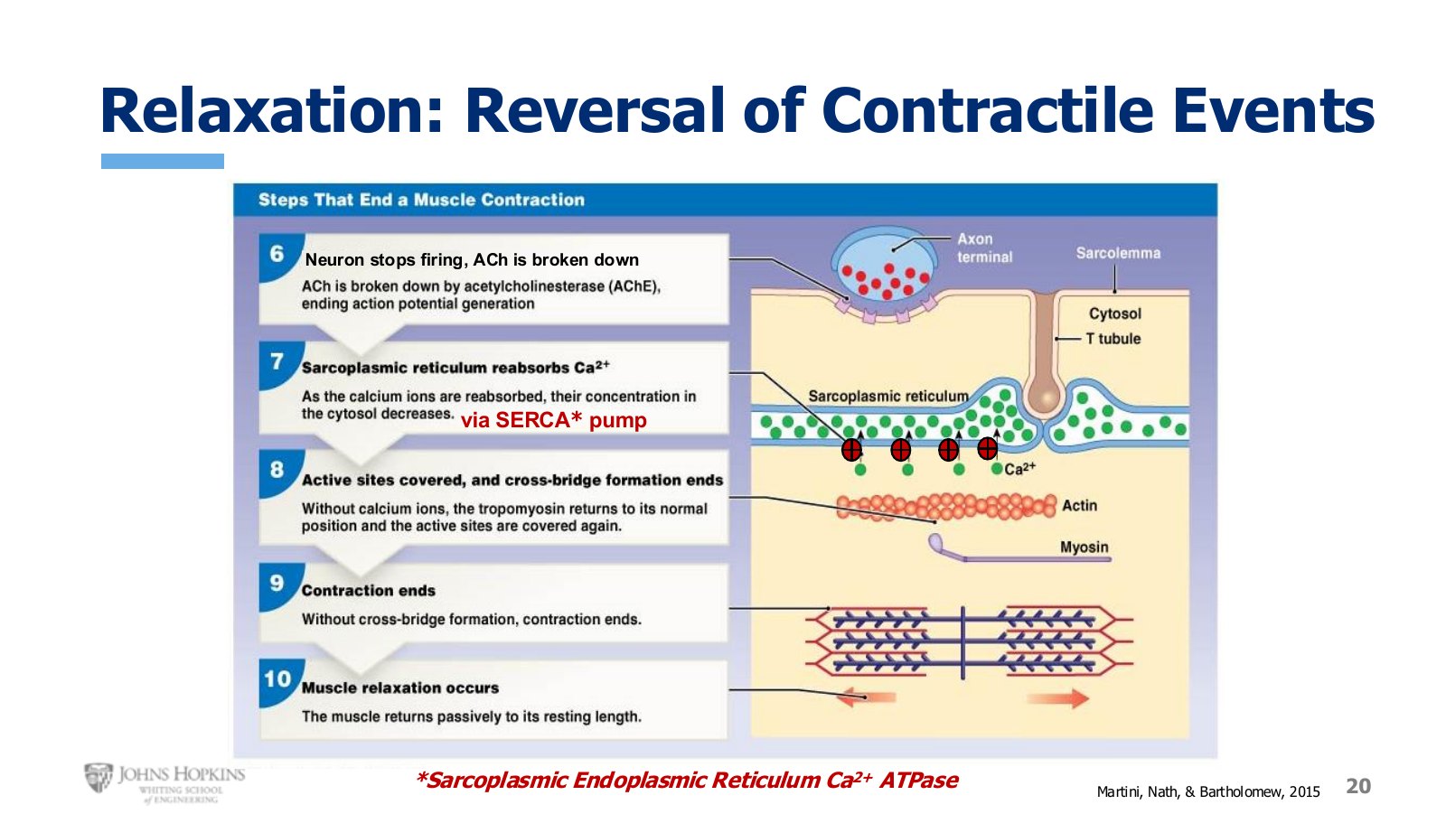
Slide 20. Relaxation, numbered 6 through 8 because steps 1 through 5 covered contraction.
The relaxation steps are pretty much the opposite of contraction.
The neuron stops firing and stops releasing acetylcholine. Acetylcholinesterase breaks down any residual acetylcholine in the cleft. No more action potential on the sarcolemma or down the T tubules, and no more calcium release from the sarcoplasmic reticulum.
Instead of releasing calcium, the sarcoplasmic reticulum gathers it back in, transporting calcium up its gradient and decreasing free calcium inside the cell. This is calcium reuptake, driven by the SERCA pump, short for sarcoplasmic endoplasmic reticulum calcium ATPase. SERCA pumps appear again in cardiac muscle relaxation later this semester.
With less free calcium available, troponin C releases it, tropomyosin covers the binding sites on actin again, and cross-bridge formation ends. Without cross-bridges there is not enough force. Contraction ends and the muscle passively returns to its resting length and tension.
A tangent worth taking: LeChatelier’s principle
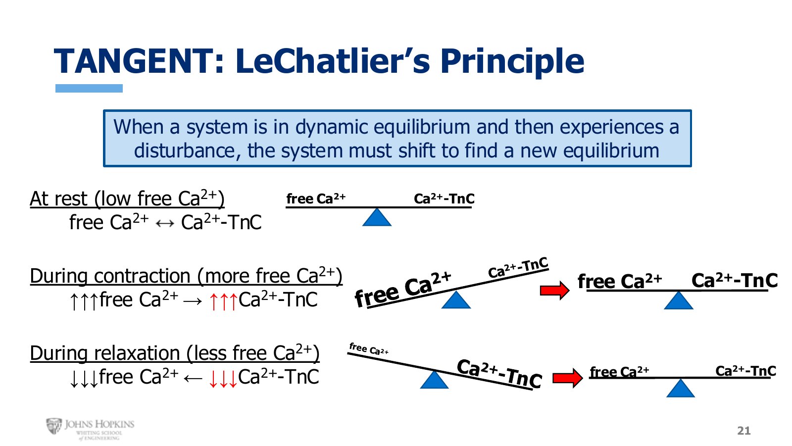
Slide 21. Free calcium and calcium bound to troponin C, shifting to find a new equilibrium three times over.
LeChatelier’s principle states that when a system in equilibrium experiences a disturbance, the system shifts and adjusts to establish a new equilibrium. The equilibrium here is the balance between free-floating calcium and calcium bound to troponin C.
At rest, intracellular calcium is quite low, but even at those levels free and bound calcium sit in equilibrium.
During contraction, the ryanodine receptor opens and free calcium rises suddenly and steeply, throwing off the balance. To restore equilibrium the reaction shifts right, pushing a lot of that new free calcium onto troponin C. A new equilibrium, at much higher levels.
During relaxation, SERCA pumps free calcium back into storage and the pool drops sharply. The reaction shifts back left, unloading calcium from troponin C, and equilibrium returns at low levels.
What excitation-contraction coupling means
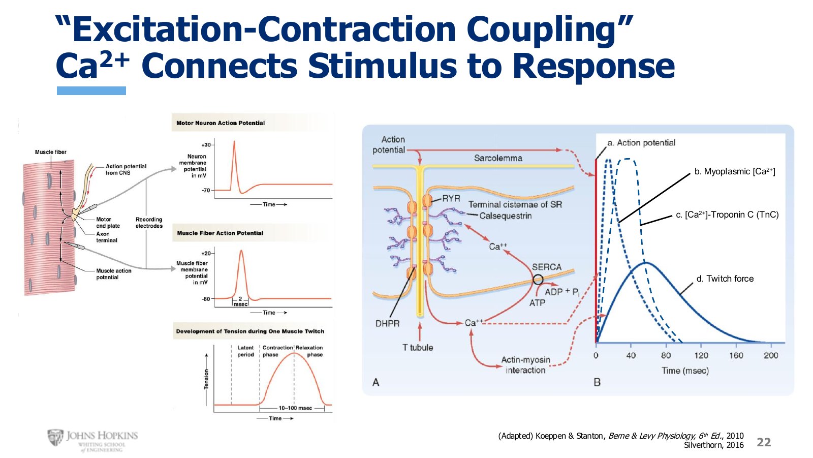
Slide 22. Muscle action potential, myoplasmic calcium, calcium bound to troponin C, and twitch force, stacked on one timescale.
Excitation-contraction coupling is the term tying together the electrical excitation of the skeletal muscle membrane with all the mechanical contractile events that follow from it. Treat the action potential on the sarcolemma as the stimulus and the resulting contraction as the response; the term highlights the cause-effect nature of how an action potential leads to muscular contraction.
Note the timescales. Action potentials are very fast, only 1 to 2 milliseconds. Twitch contractions vary in length but are always much longer, 10 to 100 milliseconds.
Calcium is the bridge. The action potential depolarizes the T tubule membrane, which opens the sarcoplasmic reticulum and lets free calcium flood into the sarcoplasm. That rise in free calcium leads to a rise in calcium bound to troponin C. That binding is the molecular signal permitting actin-myosin cross-bridging, which produces force. Without calcium release from the sarcoplasmic reticulum there is nothing linking the electrical action potential to the physical twitch. Calcium is the vital link that couples excitation to contraction.