8A · Vascular Anatomy
Between leaving the left ventricle and returning to the heart at the right atrium, blood needs to flow through literally every tissue in the body. How are the blood vessels arranged to allow this access? To explore this, we borrow some terminology from electrical circuits, thinking about the path of blood flow like the path of electrons in a circuit as it travels through the systemic circulation from left ventricle to right atrium.
In parallel and in series
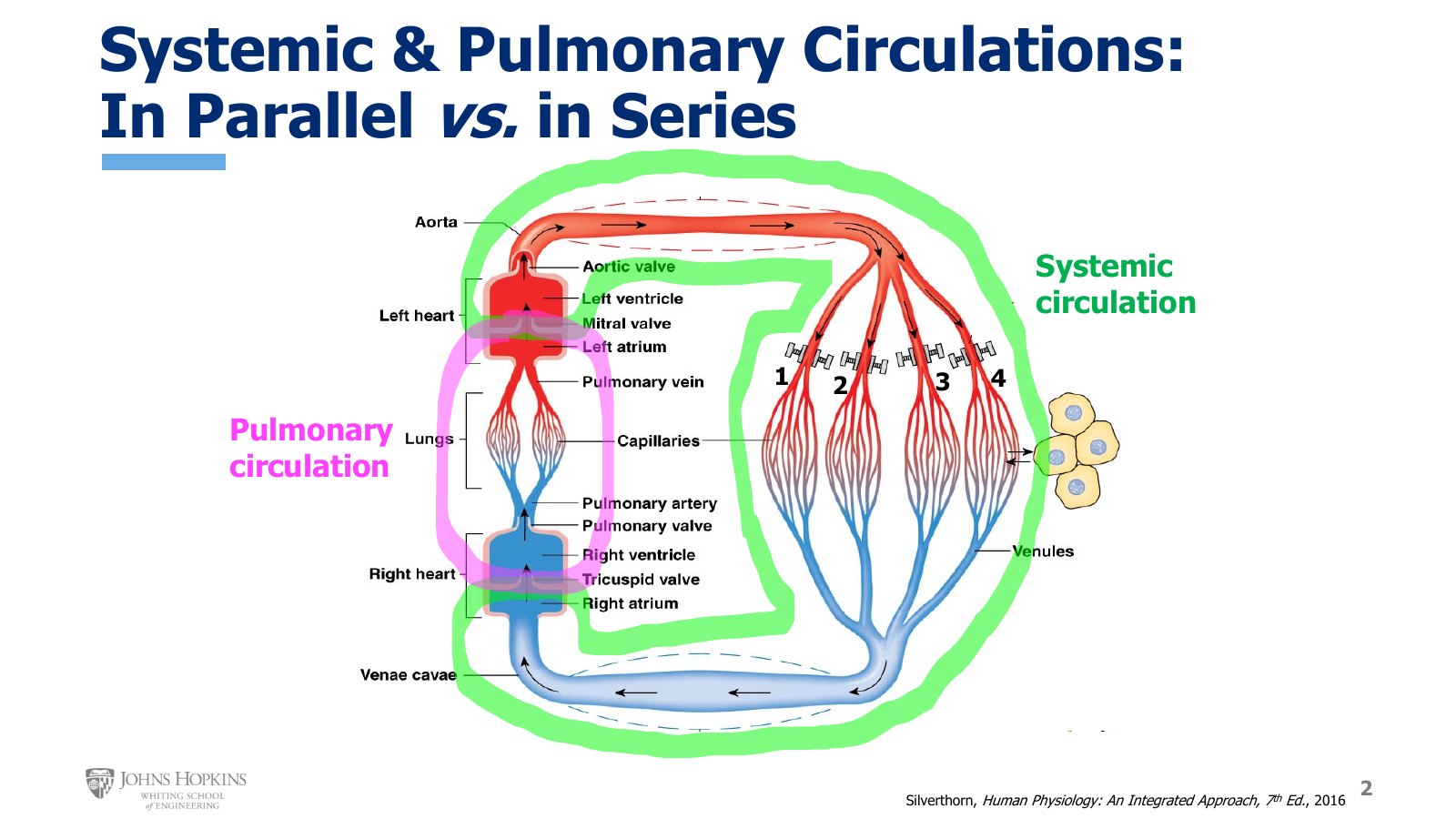
Slide 2. The systemic circulation (green) and the pulmonary circulation (pink). Capillary beds 1 to 4 each feed a separate tissue in parallel, while the two circulations sit one after the other in series.
As we distribute blood to all the tissues of the body, represented by numbered capillary beds that each feed a separate tissue, we route it through vessels arranged in parallel. That gives each tissue access to the same fresh oxygenated blood at the same blood pressure. The systemic circulation, from left ventricle to right atrium, is plumbed in parallel. The pulmonary circulation, from right ventricle to left atrium, is also plumbed in parallel.
The entire circulation, which takes in both systemic and pulmonary elements, is plumbed in series. There is a sequence here, an order, a series of vascular beds. Blood flows through the pulmonary circulation, then the systemic circulation, then pulmonary, then systemic, and so on.
The two circulations have two distinct and very different purposes, so they need to be separate, yet both need access to the entire cardiac output. The systemic circulation drops off oxygen to the tissues of the body and collects carbon dioxide as a byproduct. The pulmonary circulation drops off carbon dioxide to the lungs to exhale and picks up oxygen. Separate pumps drive each one: the right ventricle drives cardiac output through the pulmonary circulation, and the left ventricle drives cardiac output through the systemic circulation. Because of this, we don’t have to worry about any drop in circulatory pressures with the two circulations arranged in series.
Branching
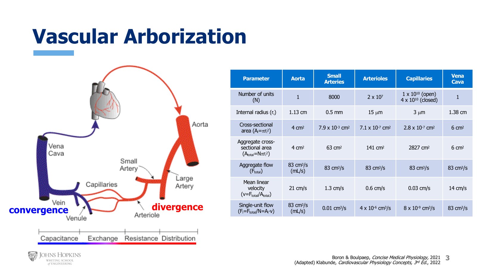
Slide 3. Divergence on the arterial side and convergence on the venous side, labeled by job: distribution, resistance, exchange, capacitance. The table compares aorta, small arteries, arterioles, capillaries, and vena cava.
The next anatomical feature to consider is the pattern of branching, or arborization, in the circulation. A lot happens between the aorta, where the left ventricle pumps, and the vena cava, which empties into the right atrium.
The arterial side is characterized by an ongoing pattern of divergence, with multiple generations of branching. With each level of branching the individual vessels get smaller, but the total number of vessels at that level increases. We go from one large aorta, to a few large arteries like the carotid, renal, and femoral arteries, to several medium-sized arteries, to lots of small arteries, to millions of arterioles.
Larger arteries simply distribute blood all over the body: to the head and arms and stomach and kidneys and legs.
Arterioles, by the millions, provide resistance and control exactly how much blood flows into the different organs and tissues. Lecture 8F covers arteriolar resistance and directing blood flow.
Capillaries do exchange: of gases, nutrients, and waste products with the tissues of the body. They are the ultimate destination of the blood in the vasculature, the whole purpose of circulating it.
Once blood has reached the capillaries, delivered oxygen and nutrients, and picked up carbon dioxide and metabolic waste products, it enters the venous side to head back to the heart. The venous side is characterized by merging, or convergence. Millions of tiny venules converge into many small veins, which merge into several medium-sized veins, then into just a few large veins, and finally into just one vena cava, which empties blood back into the right atrium.
The purpose of the veins is returning blood to the heart, and we often use the term capacitance for how veins handle blood on that return trip. Capacitance is simply another way of saying the veins serve as a blood reservoir of sorts. They can expand to hold lots of blood volume as it makes its way back to the heart. Lecture 8D treats veins as capacitance vessels in more detail.
The table on this slide is packed full of information that will help you as you consider the different levels of the circulation through this module. Keeping track of individual versus aggregate, or combined, vessel characteristics at a given level will be important going forward.
Vascular arborization, aorta to vena cava
Parameter | Aorta | Small arteries | Arterioles | Capillaries | Vena cava |
|---|---|---|---|---|---|
Number of vessels (N) | 1 | 8000 | 2 × 10⁷ | 1 × 10¹⁰ open, 4 × 10¹⁰ closed | 1 |
Internal radius | 1.13 cm | 0.5 mm | 15 µm | 3 µm | 1.38 cm |
Cross-sectional area, each vessel | 4 cm2 | 7.9 × 10⁻³ cm2 | 7.1 × 10⁻⁷ cm2 | 2.8 × 10⁻⁷ cm2 | 6 cm2 |
Aggregate cross-sectional area | 4 cm2 | 63 cm2 | 141 cm2 | 2827 cm2 | 6 cm2 |
Aggregate flow | 83 cm3/s | 83 cm3/s | 83 cm3/s | 83 cm3/s | 83 cm3/s |
Mean linear velocity | 21 cm/s | 1.3 cm/s | 0.6 cm/s | 0.03 cm/s | 14 cm/s |
Flow through each vessel | 83 cm3/s | 0.01 cm3/s | 4 × 10⁻⁶ cm3/s | 8 × 10⁻⁹ cm3/s | 83 cm3/s |
Layers of the vessel wall
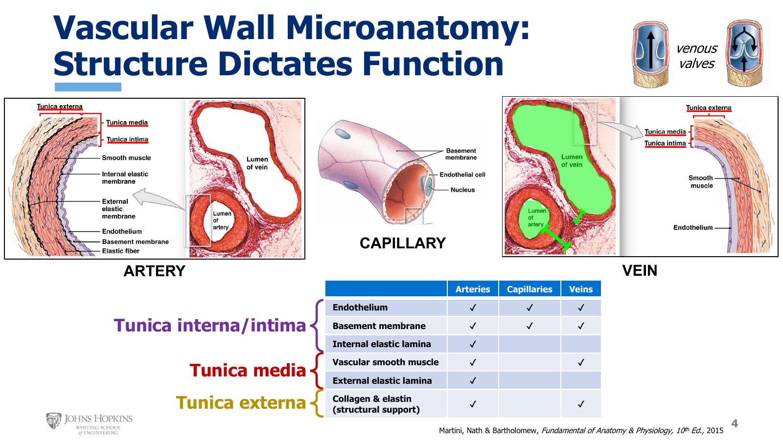
Slide 4. Artery, capillary, and vein in section, with the summary table of which wall layers each vessel has. Venous valves at top right.
Structure dictates function, and that is especially true in the vasculature. The walls of arteries and veins have three major layers, or tunicas:
Tunica intima, incredibly thin, purple here, closest to the lumen of the vessel.
Tunica media, the middle layer, red here, in the middle of the vessel wall.
Tunica externa, the external layer, tan here, the outermost layer of the wall.
Tunica intima. In the artery it has three sublayers. The endothelium is a delicate single layer of thin flat cells that fit together like puzzle pieces to line the lumen. It is easily damaged, because of its thinness and its direct exposure to the mechanical stresses of blood flow, and endothelial damage is almost universally the first step in the development of cardiovascular disease. The endothelium is anchored to the lighter purple basement membrane, which helps hold all the endothelial cells together and keeps them from floating off into the bloodstream. The black wavy line is the internal elastic membrane, a layer of elastic protein fibers that gives arteries some of their elasticity.
Tunica media. Two sublayers: the vascular smooth muscle and the external elastic membrane. The smooth muscle is what lets blood vessels change their diameter to accommodate or dictate different flow rates, by contracting or relaxing. Arteries tend to have a lot of smooth muscle, a thick tunica media. In larger arteries we often find smaller individual elastic fibers interspersed among the smooth muscle cells. The external elastic membrane gives another clearly delineated layer of elastic tissue, further contributing to the elastic property of arteries.
Tunica externa. Mostly collagen protein fibers, with a few elastic fibers woven in here and there, providing the structural support that helps vessels keep their shape.
Veins. Veins are a lot like arteries. They also have a tunica intima, media, and externa, with an endothelium, vascular smooth muscle, and collagen fibers. What they don’t have is lots of elastic tissue. There are no internal or external elastic membranes in the walls of veins. Their elastic fibers are mostly in the tunica externa, and aren’t nearly as organized or as abundant. As later lectures show, veins don’t need elasticity nearly as much as arteries do, so this really isn’t a problem.
Veins also differ in their dimensions. At a given level of branching, arteries have much thicker walls than their venous counterparts, due mostly to a much thicker layer of smooth muscle, while veins have much larger lumens than their arterial counterparts. Veins also have one-way valves, very similar in structure to heart valves, that help blood travel up toward the heart and prevent it from flowing backward, particularly when we’re standing up and blood wants to follow gravity back down to our feet.
Capillaries. All molecular exchange goes on here, so molecules need to leave and enter the bloodstream easily, and the walls are incredibly thin: a layer of endothelial cells with just enough basement membrane to hold it all together.
Because not every layer or sublayer is found in every type of vessel, here is a summary of which elements appear in arteries, capillaries, and veins.
Layers and sublayers of the vessel wall
Layer | Sublayer | Arteries | Capillaries | Veins |
|---|---|---|---|---|
Tunica intima | Endothelium | Yes | Yes | Yes |
Tunica intima | Basement membrane | Yes | Yes | Yes |
Tunica intima | Internal elastic membrane | Yes | No | No |
Tunica media | Vascular smooth muscle | Yes | No | Yes |
Tunica media | External elastic membrane | Yes | No | No |
Tunica externa | Collagen and elastic fibers, structural support | Yes | No | Yes |
Size and tissue makeup, vessel by vessel
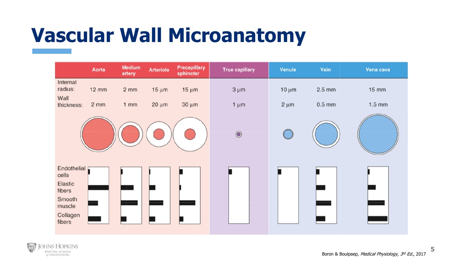
Slide 5. Internal radius, wall thickness, and relative amounts of endothelium, elastic fibers, smooth muscle, and collagen from the aorta to the vena cava.
This summary figure keeps in mind the size, dimensions, and tissue makeup of the wall for every type of artery, capillary, and vein. The internal radius of the aorta is about 12 mm, for an internal diameter of 24 mm, with a wall thickness of 2 mm. The aorta has an endothelium, lots of elastic tissue, and only a modest amount of smooth muscle, the least of any vessel here besides capillaries and venules. It has fairly significant collagen content, the most of any vessel except the vena cava.
Every single vessel in the body has an endothelium. All other components of the wall vary significantly between types and sizes of vessel, and the relative abundance of elastic fibers versus smooth muscle versus collagen fibers ends up dictating how each vessel functions in the overall circulation.
Elasticity is critical in the aorta.
Smooth muscle, and the capacity to contract or relax the vessel, is critical in the smaller arteries and arterioles.
Collagen is more abundant in the larger vessels, aorta and vena cava, to help reinforce those walls because those vessels are so large.
Vessel dimensions from the figure
Vessel | Internal radius | Wall thickness |
|---|---|---|
Aorta | 12 mm | 2 mm |
Medium artery | 2 mm | 1 mm |
Arteriole | 15 µm | 20 µm |
Precapillary sphincter | 15 µm | 30 µm |
True capillary | 3 µm | 1 µm |
Venule | 10 µm | 2 µm |
Vein | 2.5 mm | 0.5 mm |
Vena cava | 15 mm | 1.5 mm |
Regional circulations
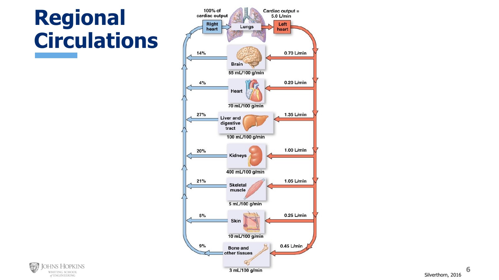
Slide 6. A resting cardiac output of 5.0 L/min divided among the regional circulations, with the flow per 100 g of tissue under each organ.
We group tissues into regions of blood flow based on how blood flow is regulated and used in that region. These are functional designations. We typically divide the body into six major circulatory regions, or vascular beds, plus a seventh catch-all group of every body tissue not yet mentioned: bone, fat, glandular tissue, stuff like that.
Each bed’s flow is its share of resting cardiac output. For the cerebral circulation:
Resting distribution of cardiac output
Circulation | Feeds | Share of resting CO | Flow | Flow per 100 g tissue |
|---|---|---|---|---|
Cerebral | Brain | 14% | 700 mL/min | 55 mL/100 g/min |
Coronary | Cardiac muscle, the heart | 4% | 200 mL/min | 70 mL/100 g/min |
Splanchnic | Gut: liver, intestines, stomach | 27% | 1,350 mL/min | 100 mL/100 g/min |
Renal | Kidneys | 20% | 1,000 mL/min | 400 mL/100 g/min |
Skeletal muscle | Skeletal muscle | 21% | 1,050 mL/min | 5 mL/100 g/min |
Cutaneous | Skin | 5% | 250 mL/min | 10 mL/100 g/min |
Everything else | Bone, fat, glandular tissue | 9% | 450 mL/min | 3 mL/100 g/min |
Only three vascular beds receive almost 70% of resting cardiac output: the gut, the kidneys, and skeletal muscle (27% + 20% + 21% = 68%). Skeletal muscle is just really abundant in the body. We might have 20 kg or more of it, so its high flow rate simply reflects that we have a lot of skeletal muscle to feed even at rest.
The gut and kidneys should get your attention. They are grossly overperfused relative to their size, at 100 and 400 mL per 100 g of tissue per minute, and grossly overperfused relative to their resting metabolic needs. So why do they get so much blood flow at rest if they don’t really need it? Because these two circulations are responsible for cleaning, filtering, and maintaining the composition of the blood. At rest, when no particular tissue has a very high demand for blood flow, we prioritize sending blood to the liver and the kidneys.
Once we go out and actually do things, walk, run, get a little hot and sweaty, different tissues start to demand more blood flow.
Exercise. Cardiac output goes way up, and blood flow to skeletal muscle and heart muscle increases dramatically at the expense of flow to the liver, kidneys, skin, and other tissues.
Heat. Cardiac output goes up, and blood flow to the skin increases dramatically at the expense of flow to the liver, kidneys, skeletal muscle, and other tissues.
Blood flow to the different regional beds is flexible, and can be adjusted to meet our needs in the moment.