8G · Capillary Function
This lecture covers capillary function and the dynamics of solute and fluid movement into and out of the capillaries.
Introduction to capillaries
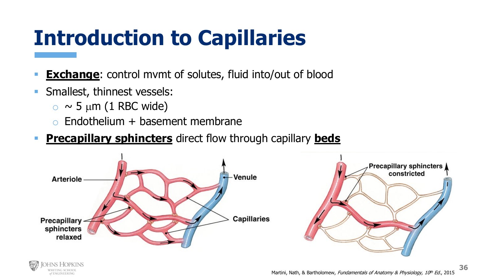
Slide 36. A capillary bed fed by an arteriole and drained by a venule. Left, precapillary sphincters relaxed and the whole bed perfused. Right, precapillary sphincters constricted and the capillaries left unperfused.
The job of the capillaries is exchange: enabling exchange of solute and water molecules with the tissues of the body. Capillaries are the smallest, thinnest blood vessels we have. They average around 5 µm in diameter, just barely accommodating the width of a single red blood cell. Their walls are incredibly thin, a layer of endothelial cells with just enough basement membrane to hold the cells together, because a thin wall makes for quick exchange of nutrients and waste products.
Blood flow through networks of capillaries, called capillary beds, is controlled directly by contraction or relaxation of precapillary sphincters, regions of smooth muscle at the entrance into the beds. In the figure, incoming red oxygenated arterial blood flows in through an arteriole. A metarteriole branches off the arteriole, and capillaries branch off both the metarteriole and the arteriole. At each entrance into the capillary bed sits a precapillary sphincter. On the left all the sphincters are open, so all the capillaries are perfused. On the right all the sphincters are closed, so there is no blood flow through the capillary bed.
Three types of capillaries
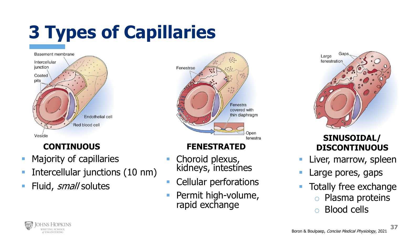
Slide 37. Continuous capillaries with only intercellular junctions, fenestrated capillaries with perforations through the endothelial cells, and sinusoidal capillaries with large fenestrations and full gaps between cells.
Continuous capillaries. The vast majority of capillaries in the body. The only gaps in the wall are at the junctions between cells, where two adjacent cell membranes meet. These intercellular junctions are only about 10 nm wide, though that is wide enough to pass solutes and fluid in and out. Not every solute can pass. Movement across these walls is limited to water molecules and small solutes like oxygen, carbon dioxide, ions, glucose, and metabolic waste products. Larger molecules, like the proteins in the plasma, cannot pass through. They are non-permeable solutes here, which will matter shortly for osmotic pressure in the blood and in the tissues.
Fenestrated capillaries. In some tissues, like the kidneys, the intestines, and the choroid plexuses in the brain that produce cerebrospinal fluid, capillaries are a bit more permeable. The name comes from the Latin fenestra, meaning window. Fenestrated capillaries have intercellular junctions just like continuous capillaries, but also small perforations, or fenestrations, holes passing through the middle of the endothelial cells, an additional avenue across the wall. The fenestrations are small enough that they don’t necessarily let larger molecules through; plasma proteins can’t get through these tiny cellular holes. But they allow a higher volume of exchange. With more pathways across the wall, more molecules can be exchanged in a given period of time, and in organs like the kidneys, intestines, and choroid plexuses it is very important to exchange lots of molecules across the capillary wall quickly.
Sinusoidal, or discontinuous, capillaries. Found in only a very few select places, such as the liver, bone marrow, and spleen. They still have intercellular junctions, but also very large fenestrations through the middle of the cells and full-blown gaps between endothelial cells. They are so permeable to pretty much everything that exchange across their walls is totally free. Their purpose is to let very large particles, like plasma proteins and blood cells, cross the wall.
Liver. It synthesizes the vast majority of our plasma proteins, so it makes sense that its capillaries are porous and permeable enough to let those proteins into the bloodstream.
Bone marrow. Blood cells are born here, and sinusoidal capillaries let newly formed blood cells enter the bloodstream.
Spleen. Blood cells are retired and recycled here, and sinusoidal capillaries let old, damaged blood cells leave the bloodstream to be broken down and recycled.
The three capillary types
Type | Where | Pathways across the wall | What crosses |
|---|---|---|---|
Continuous | Most of the body | Intercellular junctions, ~10 nm | Water and small solutes. Not plasma proteins |
Fenestrated | Kidneys, intestines, choroid plexuses | Junctions plus small fenestrations through the cells | Same small molecules, in much higher volume. Not plasma proteins |
Sinusoidal (discontinuous) | Liver, bone marrow, spleen | Junctions, large fenestrations, and full gaps between cells | Everything, including plasma proteins and blood cells |
How molecules cross the wall
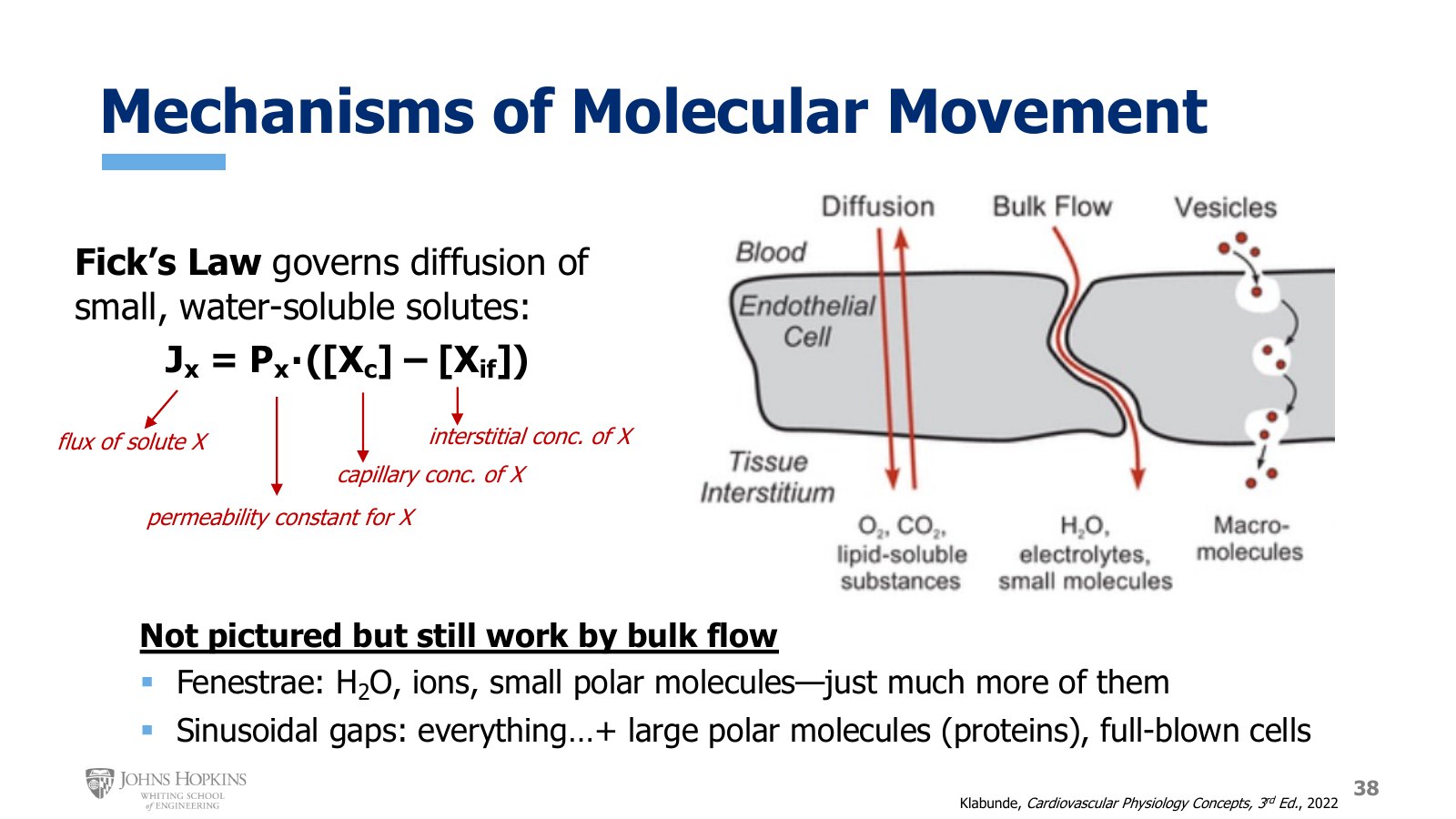
Slide 38. Diffusion of O₂, CO₂, and lipid-soluble substances through the endothelial cell, bulk flow of water, electrolytes, and small molecules between cells, and vesicles carrying macromolecules. Fick’s law annotated term by term.
We have several mechanisms for transporting molecules across the capillary wall.
Paracellular, by bulk flow. Through the intercellular junctions, molecules, especially water, ions, and small polar molecules, pass between cells, carried by bulk flow. Fenestrations aren’t pictured, but they also use bulk flow to move water, ions, and small polar molecules. The sinusoidal gaps work by bulk flow too, just without any selectivity for size or chemical characteristics.
Transcellular. There is also considerable transcellular transport of solutes, which cross the wall by passing through one side of the cell membrane, diffusing through the endothelial cell cytoplasm, and passing through the other side.
Nonpolar solutes like oxygen, carbon dioxide, and other lipophilic substances pass right through the cell membrane on their own. Remember how hydrophilic and hydrophobic substances interact with the cell membrane from the start of the semester.
Larger molecules like proteins need vesicles, undergoing endocytosis, diffusion, and exocytosis to leave the capillaries. Vesicle transport is slow and energy intensive, so not many proteins cross the capillary wall this way. That is why most capillaries are functionally impermeable to proteins.
Fick’s law
To quantify and model how solutes cross the capillary wall, Fick’s law governs diffusion of small, water-soluble molecules.
Jx is the flux, or flow, of solute X.
Px is a permeability constant for the capillary wall’s permeability to that particular solute. It changes for different solutes.
[Xc] is the concentration of solute X in the capillary.
[Xif] is the concentration of solute X in the interstitial fluid.
Flow of a solute across the capillary wall depends on the strength of the concentration gradient times the wall’s permeability to that solute.
Hydrostatic movement of water
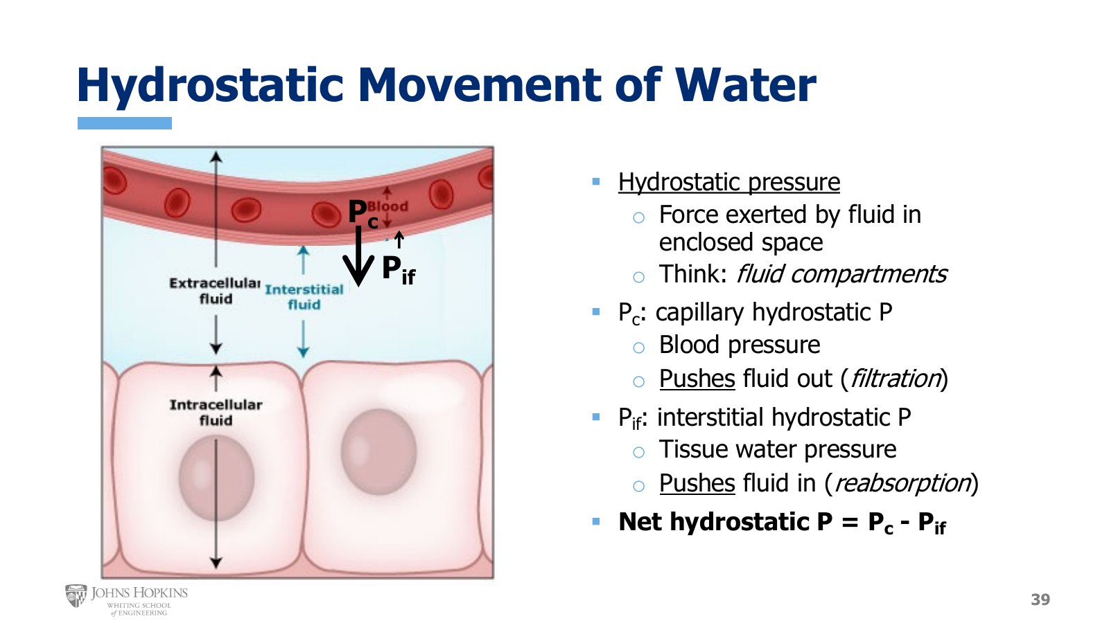
Slide 39. The capillary, interstitial fluid, and cells. A large arrow for capillary hydrostatic pressure pushes water out of the capillary, and a small arrow for interstitial hydrostatic pressure pushes it back in.
That covers solutes. Water molecules follow a different set of forces, and the first is the hydrostatic pressure gradient. In its simplest definition, hydrostatic pressure is the force exerted by a fluid in an enclosed space. In the body, the enclosed spaces where fluid can reside are the fluid compartments: the intracellular fluid compartment, the extracellular interstitial fluid compartment, and the extracellular vascular compartment.
We know from early in the semester that the osmotic gradient across the cell membrane dictates water movement between the intracellular and interstitial compartments. Right now the concern is the forces that move water between the vascular and interstitial compartments, so we examine the hydrostatic pressure in each.
Capillary hydrostatic pressure, Pc. Otherwise known as blood pressure inside the capillaries. It tends to push fluid out of the capillary and into the interstitial space, and as the size of the arrow shows, it is a strong force. Movement of water out of the capillary and into the interstitium is called filtration.
Interstitial fluid hydrostatic pressure, Pif. Essentially the pressure of the surrounding tissues and watery interstitium pressing in on the capillary, pushing water into it. Movement of water into the capillary is called reabsorption. As the small arrow shows, interstitial hydrostatic pressure is typically quite low.
The overall hydrostatic effect on water is the net hydrostatic pressure:
Osmotic movement of water
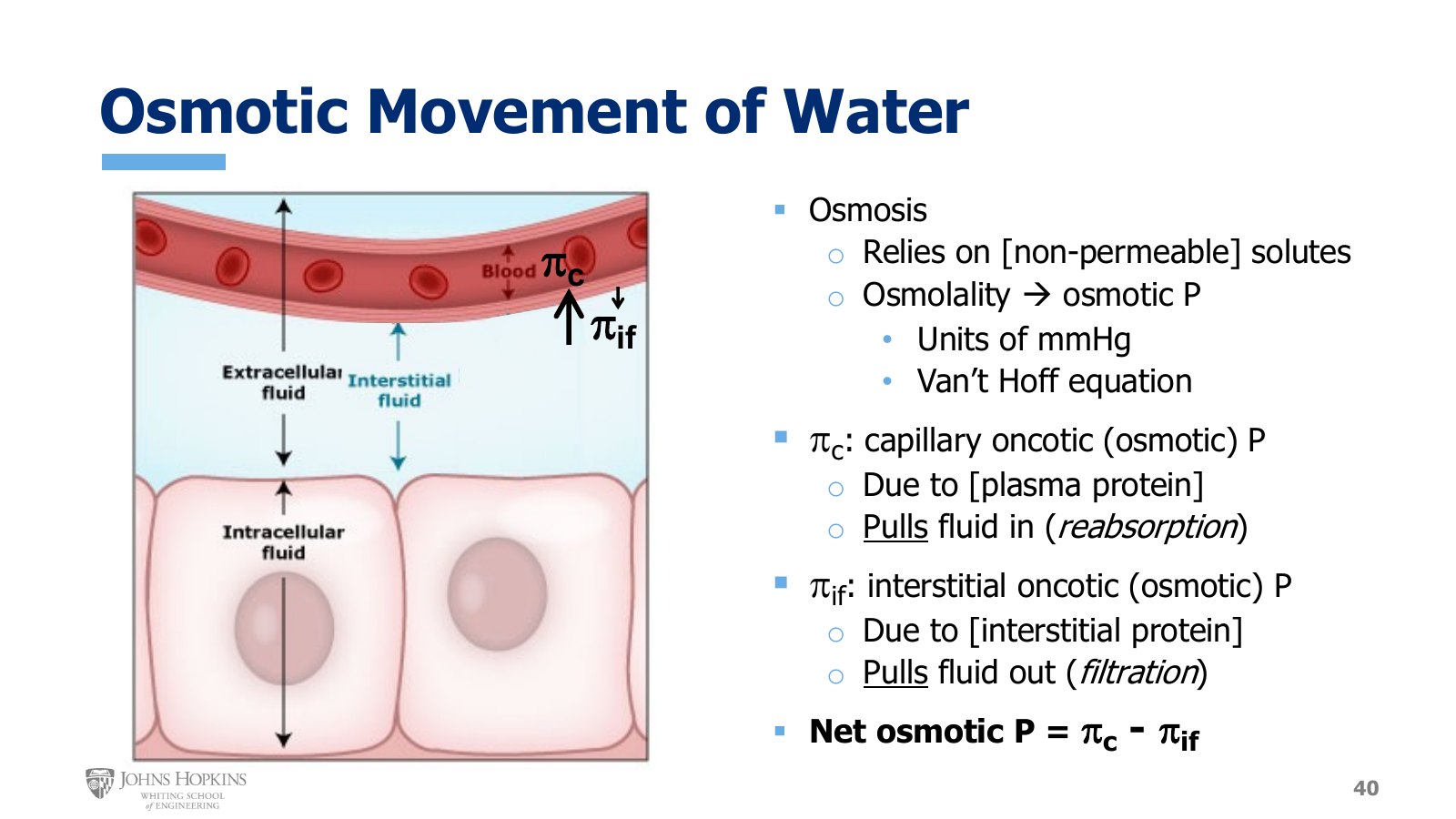
Slide 40. The same capillary with the osmotic forces. A medium arrow for capillary oncotic pressure pulls water into the capillary, and a small arrow for interstitial oncotic pressure pulls it out.
The other major force dictating water movement is osmotic pressure. From early in the semester, osmosis relies on the concentration of solutes, specifically non-permeable solutes. We can express a solution’s osmotic strength as osmolality, and we can also convert osmolality to osmotic pressure, in pressure units of millimeters of mercury, using the Van’t Hoff equation from Module 1. For transcapillary water movement we need to think in osmotic pressure, because we will soon combine osmotic and hydrostatic pressure to determine water movement, and that requires pressure units.
Capillary oncotic, or osmotic, pressure, πc. Produced by the concentration of non-permeable protein molecules dissolved in the blood. Recall that most capillary walls are impermeable to proteins. Osmotic pressures tend to draw fluid toward the source of the pressure, so osmotic pressure “pulls” water in. Capillary osmotic pressure originates in the capillaries, so it tends to pull water into them, inducing reabsorption. The medium-sized arrow shows a moderately strong force.
Interstitial fluid osmotic, or oncotic, pressure, πif. Produced by the concentration of non-permeable protein molecules dissolved in the interstitial fluid. It originates in the interstitial fluid, so it tends to pull water out of the capillaries, inducing filtration. The small arrow shows that it is quite weak. There just aren’t very many protein molecules floating around in the interstitial fluid.
The overall osmotic effect on water is the net osmotic pressure:
The four pressures acting on water across the capillary wall
Pressure | Source | Effect on water | Strength |
|---|---|---|---|
Capillary hydrostatic (Pc) | Blood pressure in the capillary | Pushes out: filtration | Strong |
Interstitial hydrostatic (Pif) | Tissues and interstitium pressing in | Pushes in: reabsorption | Quite low |
Capillary oncotic (πc) | Non-permeable plasma proteins | Pulls in: reabsorption | Moderately strong |
Interstitial oncotic (πif) | The few interstitial proteins | Pulls out: filtration | Quite weak |
Putting it all together
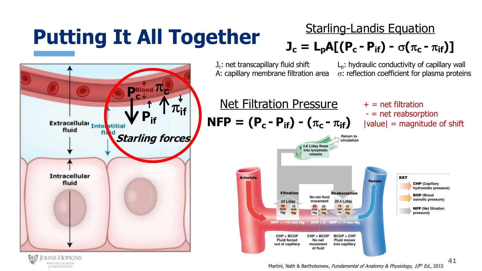
Slide 41. The four Starling forces, the Starling-Landis equation with its terms defined, net filtration pressure, and a capillary filtering at the arterial end (35 against 25 mmHg), balanced in the middle, and reabsorbing at the venous end (18 against 25 mmHg).
Together these pressures are known as the Starling forces. The Starling-Landis equation describes how net hydrostatic and net osmotic pressures work together to model overall fluid movement into or out of the capillary.
Jc is the net fluid shift across the capillary wall.
Lp is the hydraulic conductivity, or leakiness, of the capillary wall: its ability to let water across.
A is the surface area available for filtration within a capillary bed.
σ is a protein reflection coefficient that weights the net osmotic pressure. For capillaries that never let any proteins across the wall, and so reflect all proteins away from it, σ is 1. For capillaries that let all proteins through the wall all the time, σ is 0, which eliminates the influence of any osmotic gradient that might otherwise be present.
Remove the constants, which can change for different capillary beds, and pull out just the pressure terms. That gives a pretty good, if somewhat approximated, idea of how much fluid is moving across the capillary wall and in which direction: the net filtration pressure, net hydrostatic pressure minus net osmotic pressure.
Positive: net filtration predominates, and we are losing fluid from the capillary overall.
Negative: net reabsorption predominates, and we are pulling water into the capillary overall.
Absolute value: the magnitude of fluid movement. Greater numbers mean more fluid movement, lower numbers less.
Worked example: one capillary, end to end
Fairly average capillary pressures. Interstitial pressures, both hydrostatic and osmotic, tend to be so low that in this figure they are rounded down to zero and removed from consideration, leaving capillary hydrostatic and capillary osmotic pressure.
Arterial end. Capillary hydrostatic pressure pushing water outward is 35 mmHg, and capillary osmotic pressure pulling water inward is around 25 mmHg.
Positive 10 means that at the arterial end, the pressures favor filtration. Over the course of a day we filter out about 24 L of fluid in this first part of the capillaries.
Venous end. Hydrostatic pressure has fallen to 18 mmHg, while osmotic pressure is still right around 25 mmHg.
Negative 7 means that by the venous end, the pressures now favor reabsorption, but to a lesser degree than the earlier filtration, because 7 is less than 10. Over a day we typically reabsorb only about 20.4 L of the 24 L we filtered.
So 3.6 L of fluid a day is filtered out of the capillaries but not reabsorbed.
Lymphatic flow
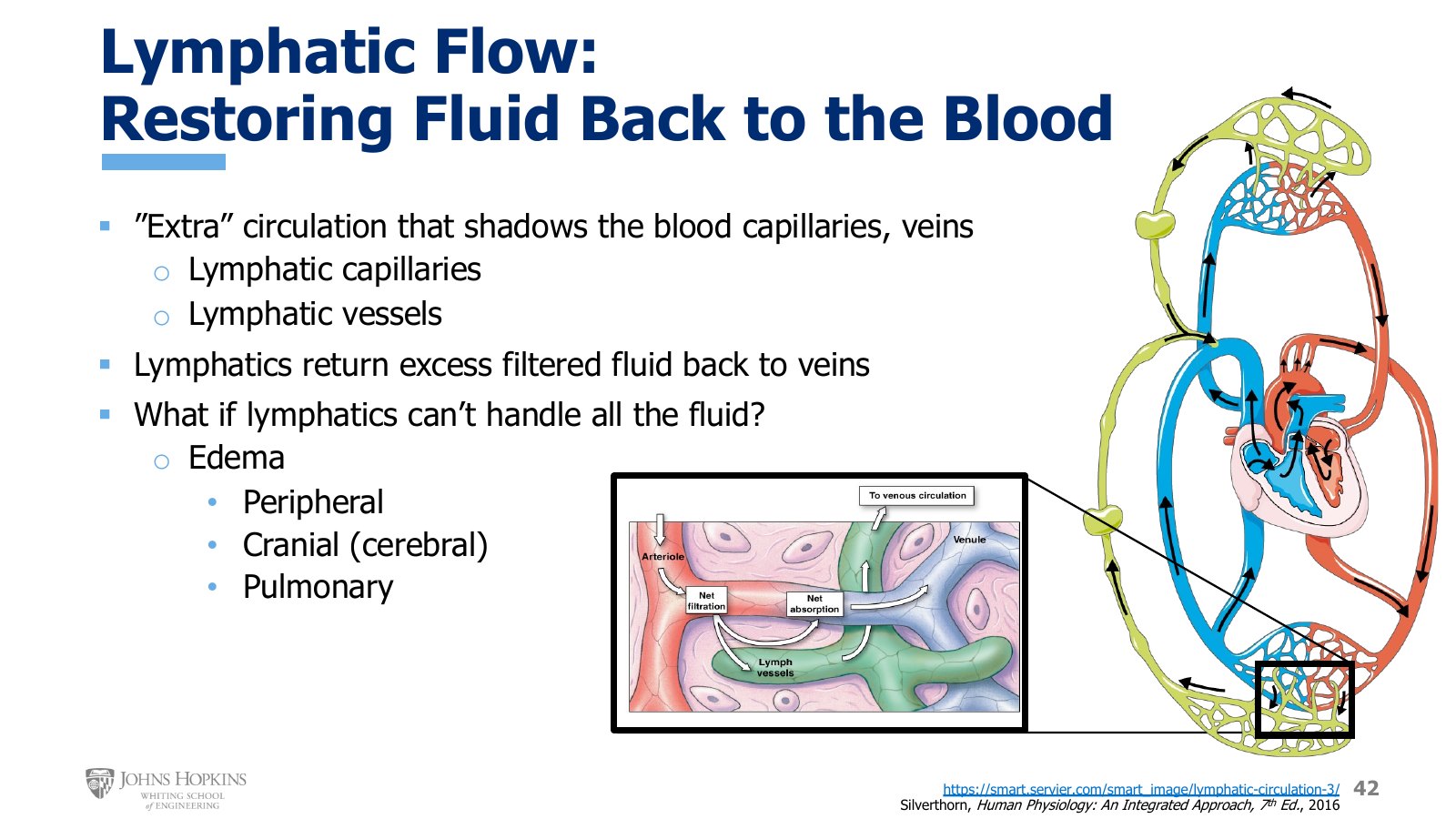
Slide 42. The lymphatic circulation (green) shadowing the blood capillaries and veins, and in the inset, lymph vessels collecting fluid filtered but not reabsorbed and returning it toward the venous circulation.
Instead of sitting in the interstitial compartment and accumulating indefinitely, the unreabsorbed fluid enters nearby lymphatic vessels. The lymphatic circulation is an additional set of vessels that shadows, or follows, the cardiovascular capillaries and veins. The lymphatic vessels are also routed through many of our lymph nodes along the way, so they play a key role in immune function too, but here we discuss only their cardiovascular application.
Lymphatic capillaries are tucked right next to blood capillaries.
Larger lymphatic vessels follow the cardiovascular veins back up toward the heart.
The lymphatic vessels empty their fluid into the cardiovascular veins near the head and neck, shortly before the veins empty into the right atrium, returning all that extra fluid to where it belongs.
The job of the lymphatic circulation, at least as it relates to capillaries, is to pick up the excess fluid that was filtered out of the cardiovascular capillaries but not reabsorbed. That way fluid doesn’t accumulate in the interstitial spaces of the body. The fluid enters the lymphatic capillaries and is carried through the lymphatic circulation until it can be returned to the blood up near the right atrium.
The same watery fluid gets a different name in each place:
In the vascular space it is plasma, and helps make up the blood.
Once it leaves the capillary and enters the interstitial space it is interstitial fluid.
Once it leaves the interstitium and enters a lymphatic capillary it is lymph.
Edema
What happens if we didn’t have lymphatic vessels, or if the volume of filtered fluid simply overwhelms the lymphatic capacity to pick it all up? We get edema, the accumulation of fluid in the interstitial space.
Peripheral edema. Familiar to most of us: the swelling of an injured knee, or the puffy ankles at the end of a long day on our feet. It may be a little uncomfortable, but it isn’t life-threatening, because our peripheral tissues are pretty soft and distensible and can accommodate quite a bit of swelling before compressing any vital structures.
Cranial, or cerebral, edema. Can be life-threatening. In the cranium, or skull, there is no room for fluid to accumulate in a tight space encased entirely in rigid bone. The fluid can’t expand outward, so it expands inward instead, compressing brain tissue.
Pulmonary edema. Can be life-threatening. In the lungs it is very important to keep fluid in its own space and air in its own space. Pulmonary edema means excess fluid starts to fill the air sacs, compromising our ability to breathe and exchange gases.