8F · Vascular Resistance (R)
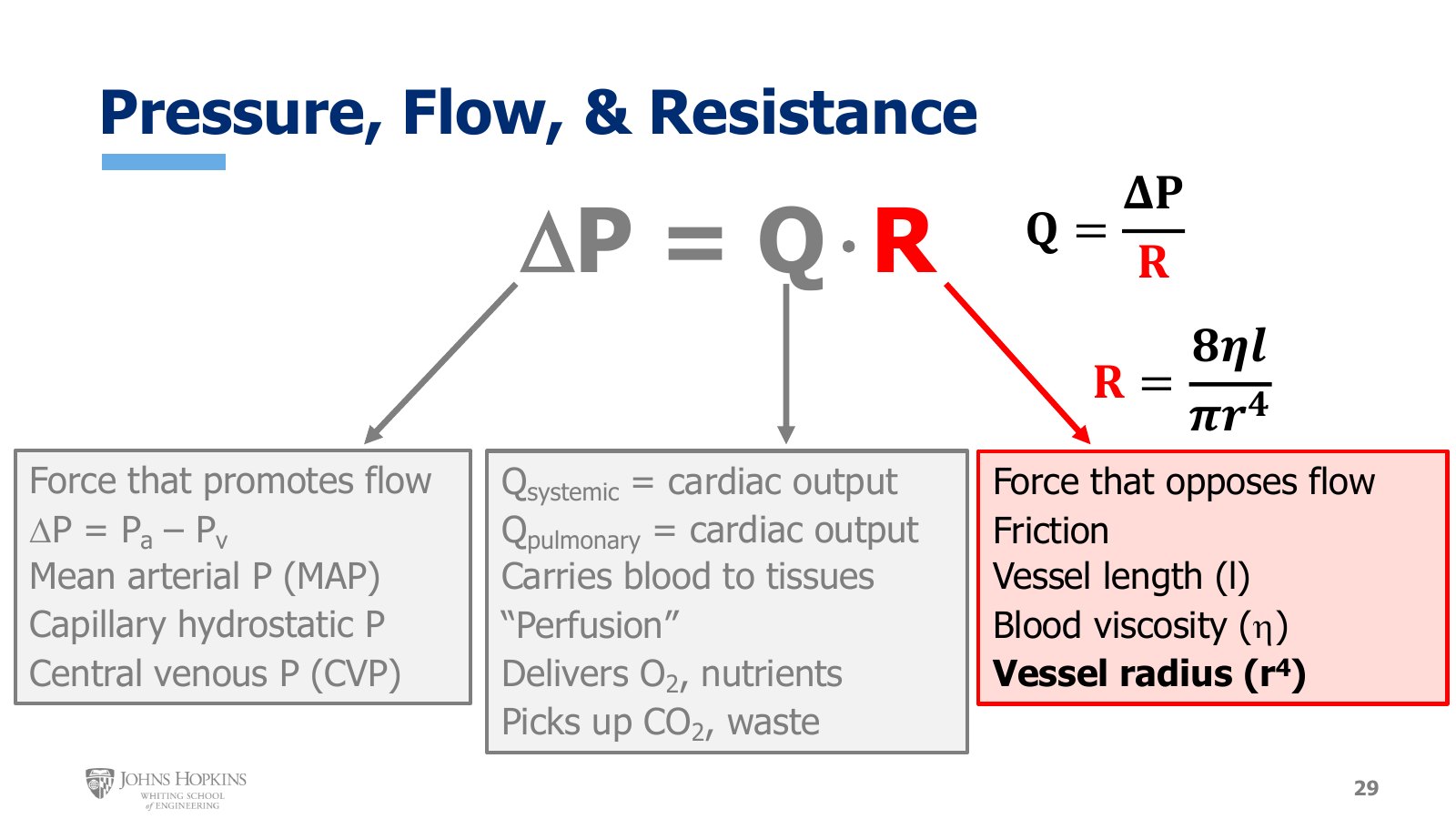
Slide 29. The overview a final time, with R highlighted beside Q = ΔP/R and R = 8ηl/πr⁴. Vessel radius is in bold.
The big picture first. Resistance is a critical factor in determining both flow and pressure. It is the force that opposes flow, and it is largely a function of friction: friction between the blood and the vessel wall, and friction between adjacent layers of blood flow. From the Hagen-Poiseuille equation, the major physiological factors affecting resistance are vessel length, blood viscosity, and vessel radius, with radius raised to the fourth power and so carrying the most mathematical weight.
What we know so far
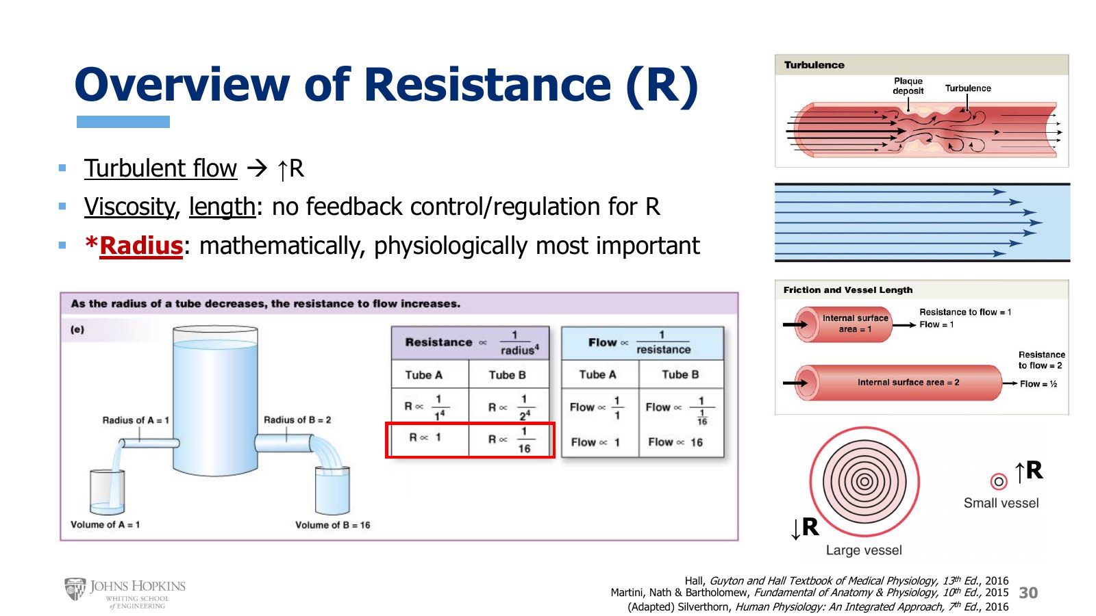
Slide 30. Turbulence past a plaque deposit, friction doubling with doubled vessel length, the tube A and tube B comparison of radius, resistance, and flow, and many laminae in a large vessel against one in a small vessel.
From the end of lecture 8E, turbulent flow patterns have high resistance, because so much of the forward kinetic energy of laminar flow gets dissipated and lost with turbulence. It is just harder to make blood flow forward, which is the definition of vascular resistance. Assuming laminar flow, we can now consider viscosity, length, and radius as predictors of resistance.
Viscosity and length
Viscosity and length are interesting. They certainly contribute to vascular resistance, but they are not under overt regulated feedback control. We don’t have mechanisms to change vessel length or blood viscosity in an attempt to regulate vascular resistance. They are simply variables that exist in the system and must therefore be considered.
Viscosity. Blood viscosity, most strongly determined by red blood cells and hematocrit, tends to oppose the forward flow of blood because it creates friction between adjacent layers of flow, and wherever there is friction, movement is more difficult. We do have mechanisms to regulate red blood cell concentration and so hematocrit, but those mechanisms are not driven by or responsive to vascular resistance. It is not a variable the body uses to adjust resistance.
Length. Vessel length contributes to resistance because it increases the vessel surface area over which friction between the blood and the vessel wall can develop. But we have no way to manipulate or regulate vessel length. It is simply a function of body growth. As we grow from infancy to childhood to adolescence and into adulthood, our vessels get longer, so resistance increases over time too. This change is very slow, but it helps explain why arterial blood pressures in babies and young children are so different from those in adults.
Radius
Radius exerts the strongest mathematical influence over the calculation of resistance, because its effect is amplified to the fourth power. In the schematic, tube B has a radius twice as large as tube A.
A twofold increase in radius results in a 16-fold decrease in resistance. Tube B has a resistance 16 times lower than tube A. Conversely, because tube A has half the radius of tube B, it has a resistance 16 times higher than tube B.
Why radius matters at all. Friction between blood and vessel wall is still a factor. In a narrow vessel, like the small one on the right, there is only one layer, or lamina, of flow, and it is in direct contact with the vessel wall. All the flow in that vessel is slowed by friction with the wall opposing forward flow: higher resistance. In the larger vessel on the left there are many adjacent layers of laminar flow. The outermost layer is still in contact with the wall and still experiences friction, but the larger radius allows many inner layers that never touch the wall and so are unaffected by friction with it. More of the blood in the larger vessel isn’t experiencing friction that opposes flow, so the larger vessel has lower resistance.
Arterioles: the resistance vessels
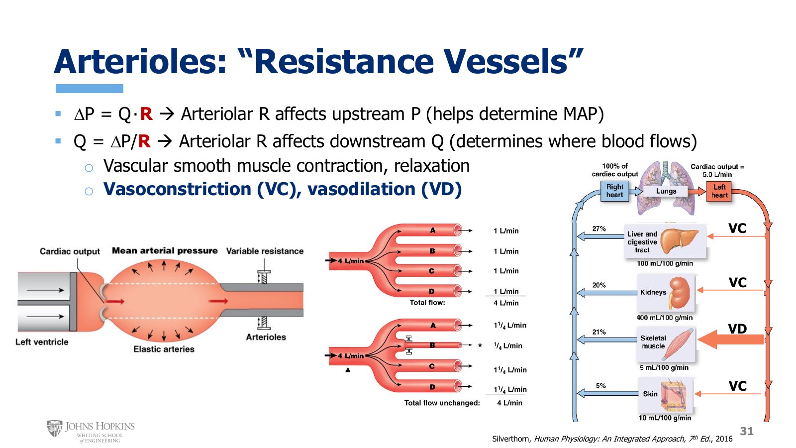
Slide 31. Arteriolar resistance sets upstream pressure and downstream flow. Center, four arterioles sharing 4 L/min before and after arteriole B constricts. Right, vasoconstriction in gut, kidney, and skin and vasodilation in skeletal muscle.
Radius is both mathematically and physiologically the most important factor. Physiologically, because we have dedicated, regulated feedback mechanisms that change vessel radius for the designated purpose of changing resistance, in order to affect flow and pressure.
The arterioles, at the end of the arterial side just before the capillaries, are known as the resistance vessels of the body. By virtue of their size, dimensions, tissue makeup, and placement in the circulation, when these vessels constrict and dilate to change their radius they exert a disproportionately large influence on vascular resistance.
When we manipulate resistance we also change flow and pressure. Contracting or relaxing arteriolar smooth muscle constricts or dilates the arteriolar radius, with effects in both directions:
Upstream. Arteriolar constriction or dilation affects pressure in the larger arteries upstream, and so influences mean arterial pressure.
Downstream. Arteriolar constriction lowers flow rates and tissue perfusion downstream of the constriction. Arteriolar dilation raises them. So constricting and dilating arterioles is a major tool for deciding which tissues receive more or less blood flow at the moment.
To keep the terminology straight: contraction and constriction of vessels is vasoconstriction, VC. Relaxation and dilation of vessels is vasodilation, VD.
Rerouting flow
In the middle image, four arterioles A, B, C, and D all have the same diameter and so the same flow rates. Upstream flow is 4 L/min, split equally among four arterioles.
Now arteriole B constricts, and flow through it falls to a quarter of a liter per minute. That forces more blood through arterioles A, C, and D.
Total flow through the arterioles is still 4 L/min, but we’ve shifted where the blood is routed.
A more physiological version uses the regional circulations from lecture 8A: the splanchnic or gut bed, the renal or kidney bed, the skeletal muscle bed, and the cutaneous or skin bed, at their original resting flows. Now say you get up and go for a run on a cool, brisk day. Your skeletal muscle now needs a lot more oxygenated blood flow than at rest, since it has to make a lot more ATP. So the arterioles in that bed vasodilate to let more blood flow in. To support the muscles’ need, the arterioles in the gut, kidneys, and skin simultaneously vasoconstrict, rerouting their blood flow to skeletal muscle, the tissue that needs it most right now.
Arterioles use vasoconstriction and vasodilation to finely tune how much flow comes through for tissue perfusion, matching each tissue’s need in the moment, while also making sure mean arterial pressure is maintained at levels adequate to keep driving perfusion.
Vasodilation: how we raise r
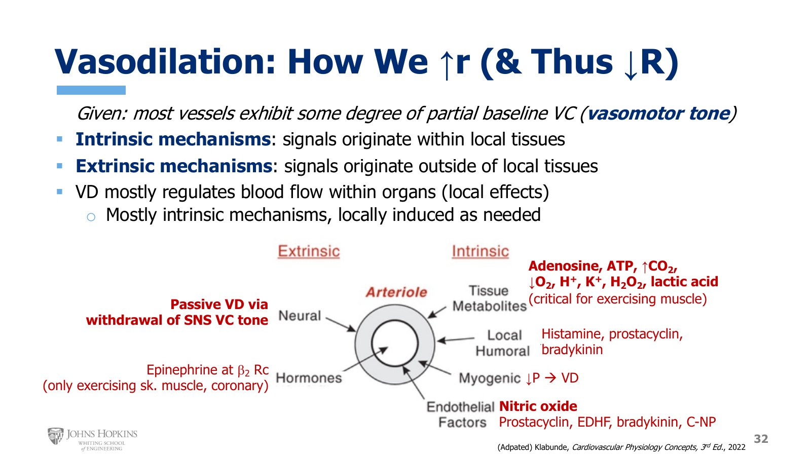
Slide 32. Signals that dilate an arteriole. Extrinsic: neural (passive dilation on withdrawal of sympathetic tone) and hormonal (epinephrine at beta-2 receptors). Intrinsic: tissue metabolites, local humoral factors, myogenic response, and endothelial factors.
Now that we know how much changes in resistance matter for upstream arterial pressure and downstream tissue perfusion, the next question is how vessels change their diameters. How do we stimulate relaxation or constriction of vascular smooth muscle to change radius? Vasodilatory mechanisms first. There are two general sources of vasodilation:
Intrinsic mechanisms use dilator signals that originate in the local tissue where the dilation needs to occur.
Extrinsic mechanisms use signals that originate outside the local tissues.
Intrinsic dilators come from four places.
The tissues that use the oxygen. Metabolic byproducts diffuse out of the cells and into the vessel wall, stimulating relaxation of vascular smooth muscle. These vasodilatory metabolites include adenosine, ATP, increased carbon dioxide, decreased oxygen, hydrogen ions, potassium ions, hydrogen peroxide, and lactic acid. Metabolic vasodilation is particularly important in exercising skeletal muscle and in the coronary vascular bed.
Nearby cells that deliberately release molecules that diffuse into the tunica media and stimulate smooth muscle relaxation. These locally released dilators include histamine, prostacyclin, and bradykinin.
The smooth muscle itself. Myogenic literally translates to muscle origin. Smooth muscle cells are sensitive to stretch and pressure, so a decrease in the stretch and pressure the smooth muscle experiences induces vasodilation.
The endothelium, which releases several powerful vasodilator molecules that diffuse into the smooth muscle: nitric oxide, prostacyclin, endothelium-derived hyperpolarizing factor (EDHF), bradykinin, and C-type natriuretic peptide (CNP).
Extrinsic dilators are a different story. In theory, extrinsic signals could come from the nervous system, or from the endocrine system releasing hormones into the bloodstream that eventually circulate to the target tissue. Because nerves and hormones travel pretty much everywhere in the body, extrinsic mechanisms typically exert whole-body effects. But we don’t have much extrinsic vasodilation.
Withdrawal of sympathetic tone. Probably the biggest contributor, and it is passive. Most of our blood vessels sit under resting conditions with some degree of sympathetically induced vasoconstriction, called resting vasomotor tone. If a decrease in sympathetic nerve activity removes that constriction, the vessel dilates a little simply because it is no longer constricting.
Epinephrine. The only other real extrinsic vasodilatory mechanism is hormonal. Epinephrine, a hormone triggered by the sympathetic nervous system, binds beta-2 receptors in skeletal muscle and coronary vessels. This mechanism is relatively minor and is only relevant during exercise.
Vasodilation is mostly driven by intrinsic mechanisms that originate within the local tissue. That means vasodilation in the body mostly exists to regulate downstream organ perfusion, engaging these local mechanisms for a localized effect only when needed.
Vasoconstriction: how we lower r
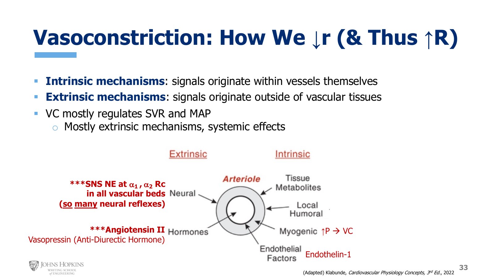
Slide 33. Signals that constrict an arteriole. Extrinsic: sympathetic norepinephrine at alpha-1 and alpha-2 receptors in all vascular beds, angiotensin II, and vasopressin. Intrinsic: the myogenic response and endothelin-1.
The general definitions of intrinsic and extrinsic mechanisms carry over.
Intrinsic vasoconstriction. We just don’t use it very much. Technically, certain vascular beds show a myogenic vasoconstriction, where an increase in vessel stretch and pressure stimulates the smooth muscle to contract and constrict. And the endothelium technically does release a vasoconstrictor molecule, endothelin-1. But we don’t really use these mechanisms very often to initiate vasoconstriction.
Extrinsic vasoconstriction across the whole body is much more common.
Sympathetic nervous system. By far the most active vasoconstrictor influence in the body, stimulating vasoconstriction on a second-to-second and minute-to-minute basis. Sympathetic neurons release norepinephrine onto vascular smooth muscle in all vascular beds of the body, and norepinephrine binds alpha-1 and alpha-2 receptors to stimulate smooth muscle contraction, as in the smooth muscle module. The sympathetic nervous system participates in many regulatory reflexes, so sympathetic vasoconstriction is being stimulated to at least some degree pretty much all the time.
Angiotensin II, a powerful vasoconstrictor hormone.
Vasopressin, also called antidiuretic hormone, also a hormone with considerable constrictor influence.
Sympathetic reflexes that affect the cardiovascular system come in a later module this semester. Angiotensin II and vasopressin come in Physiology II, with the endocrine system and hormones in depth.
Vasoconstriction is mostly driven by extrinsic mechanisms that originate outside the local tissue and stimulate whole-body, systemic vasoconstriction. That is because vasoconstriction is mostly used to increase systemic vascular resistance, SVR, with the intent of maintaining upstream mean arterial pressure.
Vasodilation against vasoconstriction
Feature | Vasodilation (raise r, lower R) | Vasoconstriction (lower r, raise R) |
|---|---|---|
Mostly driven by | Intrinsic, local mechanisms | Extrinsic, whole-body mechanisms |
Mostly used to | Regulate downstream organ perfusion | Raise SVR and maintain upstream MAP |
Main intrinsic signals | Metabolites, histamine, prostacyclin, bradykinin, myogenic (↓P), nitric oxide and other endothelial factors | Myogenic (↑P), endothelin-1. Rarely used |
Main extrinsic signals | Passive, by withdrawal of sympathetic tone. Epinephrine at beta-2, minor, exercise only | Sympathetic norepinephrine at alpha-1 and alpha-2. Angiotensin II. Vasopressin |