4A · Introduction to Smooth Muscle
Smooth muscle is utterly, completely different from skeletal muscle. There are thick and thin filaments, and calcium still drives contraction, but that is about it. It is simply a very different beast.
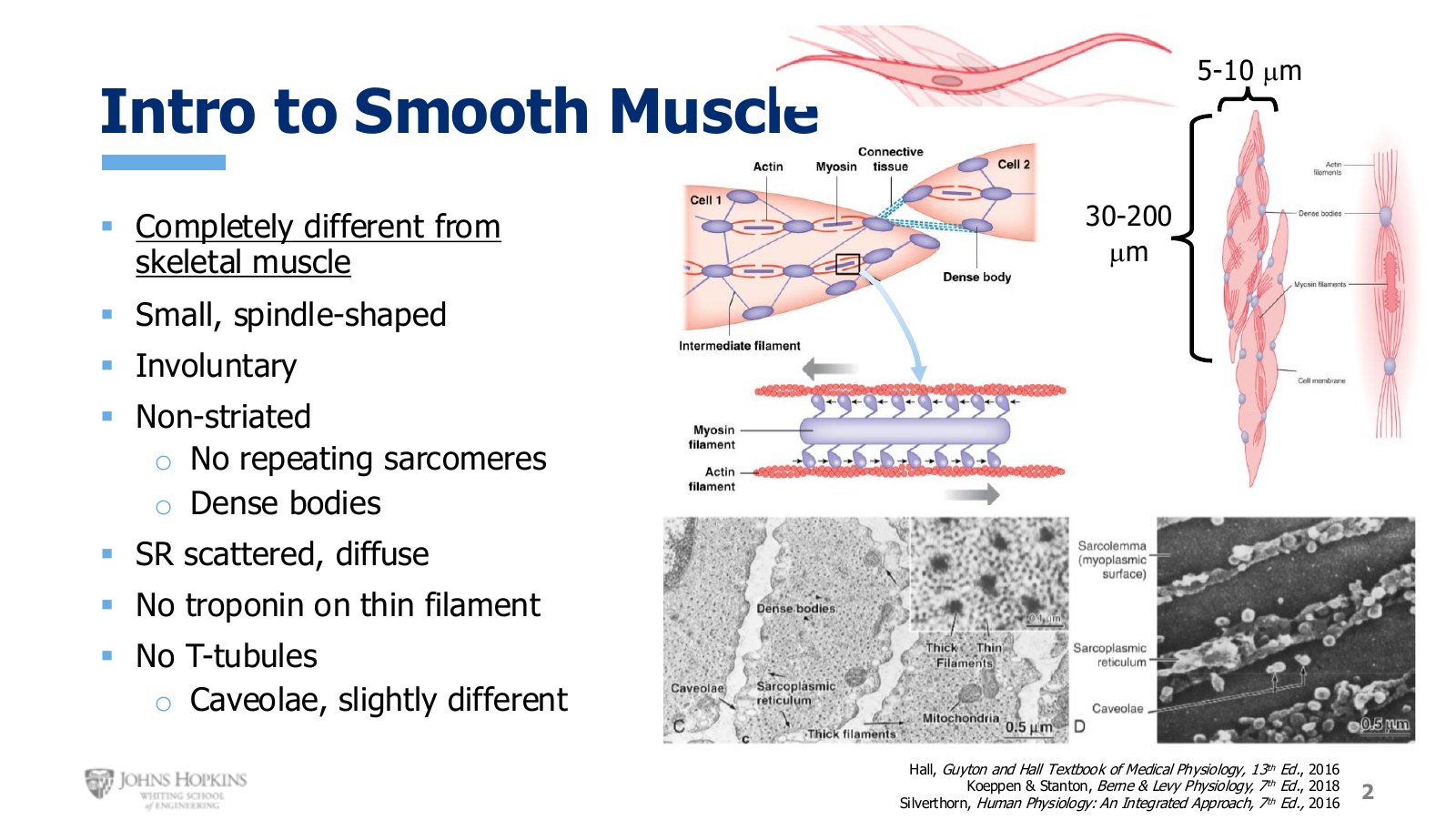
Slide 2. Smooth muscle cells beside the skeletal muscle we know. Spindle-shaped, far smaller, and with no striations.
Smooth muscle against skeletal muscle
Feature | Skeletal | Smooth |
|---|---|---|
Shape | Cylindrical | Spindle, tapered at the ends |
Thickness | ~100 µm | 5–10 µm |
Length | up to 30 cm | 30–200 µm |
Control | Voluntary | Involuntary |
Striations | Vivid | None |
Thin filament anchor | Z discs, repeating sarcomeres | Dense bodies, scattered |
Sarcoplasmic reticulum | Abundant, organized | Less abundant, scattered, diffuse |
Calcium sensor on thin filament | Troponin | None |
Membrane invaginations | T tubules | Caveolae, much shallower |
Smooth muscle is involuntary because the autonomic nerves that control it are involuntary. We have no deliberate control over making smooth muscle contract and relax, and depend entirely on unconscious reflex regulatory mechanisms.
It is non-striated because the contractile filaments are not organized into repeating sarcomeres. Instead the thin filaments anchor to structural proteins called dense bodies, scattered around the cell, with the thick filaments found between them.
There are no T tubules. Instead there are much shallower periodic indentations in the membrane called caveolae, which is where the majority of ligand receptors, membrane enzymes, and ion channels are found.
Categorizing by location
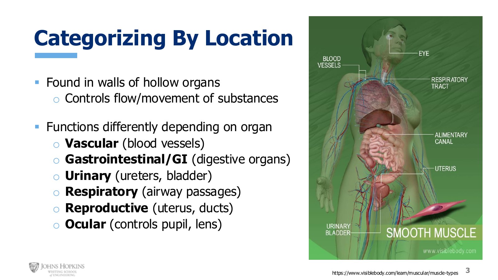
Slide 3. Smooth muscle sits in the walls of hollow organs throughout the body.
Smooth muscle is found in the walls of most hollow organs, where it controls the movement of air, liquid, and solids. It responds differently to different stimuli depending on the organ it sits in.
Vascular, in blood vessel walls. Dilates or constricts vessels, affecting blood flow to different parts of the body.
Gastrointestinal, in the hollow organs of the GI tract, also called the alimentary canal. Moves food for digestion and opens and closes sphincters.
Urinary, in the ureters moving urine from kidneys to bladder, and in the bladder wall contracting to evacuate urine.
Respiratory, in the airways. Narrows or widens them, affecting how much and how easily air flows into the lungs.
Reproductive, in the uterus, vaginal walls, and fallopian tubes, and in the vas deferens, glandular ducts, and sphincters.
Ocular, controlling pupil diameter and lens positioning.
This semester covers vascular smooth muscle under arteries and veins, and respiratory smooth muscle under the airways. Physiology II covers the gastrointestinal, urinary, reproductive, and ocular cases.
Categorizing by contraction pattern
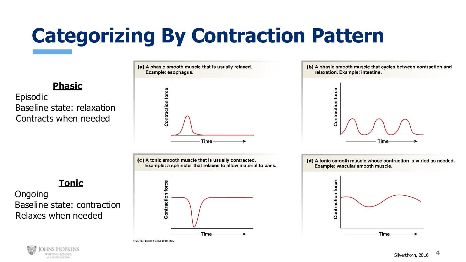
Slide 4. Phasic muscle rests and contracts when needed. Tonic muscle contracts and relaxes when needed.
Phasic contraction is episodic and intermittent. The baseline state is relaxed most of the time, and the muscle contracts only when necessary. The esophagus is truly relaxed almost all the time, producing contraction only when food or liquid is actively swallowed. The intestines contract fairly often to mix food or propel it along, but always return to a relaxed baseline.
Tonic contraction is mostly ongoing, because contraction is the baseline state and the muscle relaxes only when needed. Sphincters are the clearest example, staying closed most of the time and relaxing to open when necessary: out of the esophagus into the stomach, out of the stomach into the small intestine, out of the small intestine into the large intestine, and from the rectum to the outside world. Vascular smooth muscle is also tonic, with some contraction always necessary, but the degree varying to maintain blood pressure.
Categorizing by connectivity
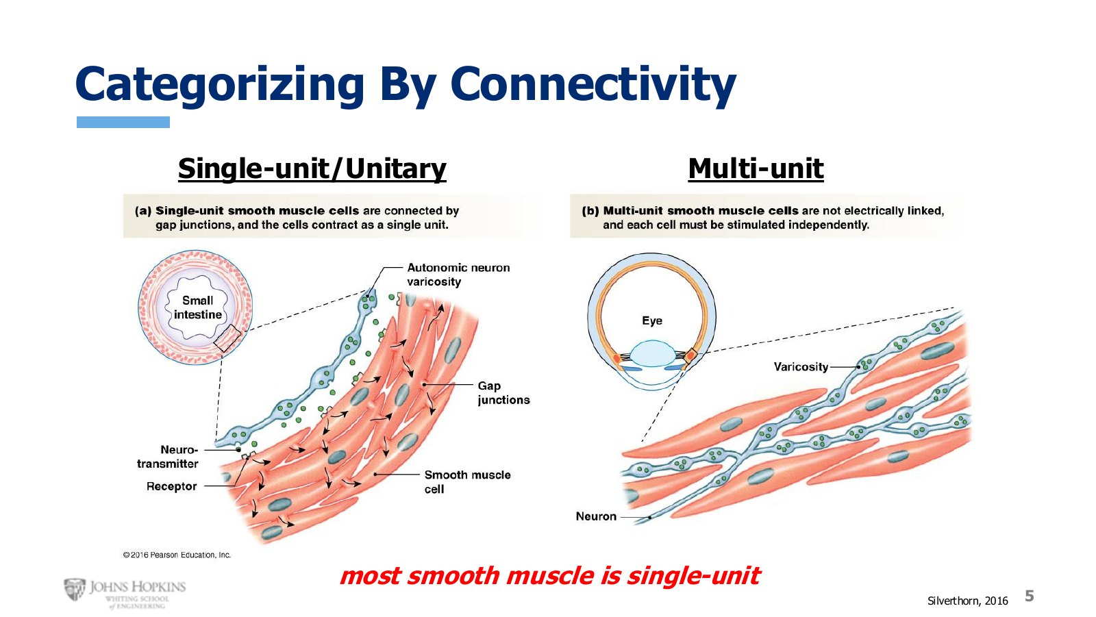
Slide 5. Single-unit cells share cytoplasm and fire together. Multi-unit cells are independent and each needs its own innervation.
In a single-unit, or unitary, arrangement the cells are all connected, sharing cytoplasm and conducting action potentials to each other. A neuron stimulating just a small number of cells is enough, because those few pass the signal to all nearby cells, letting the whole group function synchronously as one unit.
The axon terminal here differs from what innervated skeletal muscle. It carries several swellings along its length, called varicosities, that contain and release neurotransmitter. So a single neuron can contact several smooth muscle cells, and can make several points of contact with a single cell.
In multi-unit smooth muscle the cells are not electrically connected. They are independent, do not synchronize, and do not contract as one unit, so each cell needs its own source of neural stimulation through much more abundant innervation. With each cell stimulated independently there is finer control over exactly how many cells fire for a given task.
Autonomic innervation
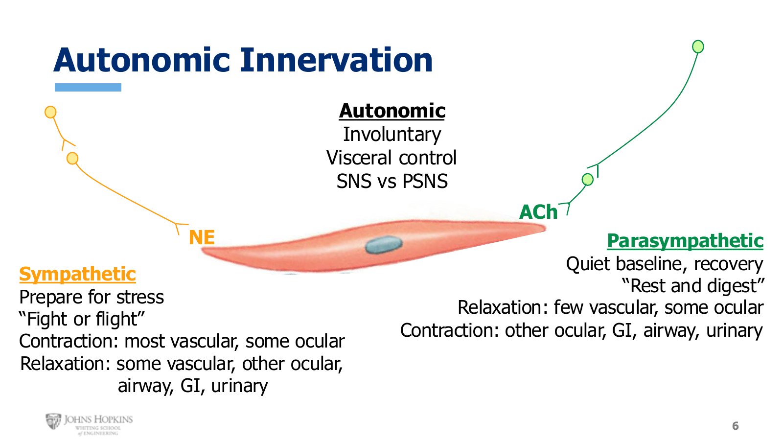
Slide 6. Sympathetic and parasympathetic effects on smooth muscle, organ by organ. Neither branch simply contracts or relaxes everything.
Where skeletal muscle was innervated by somatic motor neurons, smooth muscle is innervated by autonomic motor neurons. These are involuntary, controlling visceral organs and functions that run in the background without our awareness. The autonomic branch splits into the sympathetic nervous system, or SNS, and the parasympathetic nervous system, or PSNS.
Sympathetic. Two neurons in sequence out of the spinal cord, the first short and the second fairly long. The second releases norepinephrine. The purpose is to prepare the body for an imminent stress, the fight or flight system, whether that is exercise, a big presentation, or a major job interview.
Contract: most vascular smooth muscle, raising blood pressure so blood is pressurized enough to reach the muscles that need it. Some ocular smooth muscle, which sounds paradoxical but helps the pupils dilate to let more light in.
Relax: some vascular smooth muscle, such as in actively exercising muscles, to allow more blood flow in. Other ocular smooth muscle, also aiding pupil dilation. Respiratory smooth muscle, allowing more oxygen into the lungs. GI and urinary smooth muscle, slowing digestion, defecation, and urination, all of which would get in the way during a bicycle race.
Parasympathetic. The opposite in nearly every way, bringing us back down from a stressor to baseline, resting and digesting and recovering. Also two neurons in sequence, but this time the first is long and the second much shorter. The second terminates on smooth muscle releasing acetylcholine, binding receptors that differ from those in skeletal muscle.
Relax: some vascular smooth muscle in the male reproductive system, and some ocular smooth muscle aiding pupil constriction.
Contract: other portions of ocular smooth muscle, also aiding pupil constriction. GI and urinary smooth muscle carrying out digestion, defecation, and urination. Airway smooth muscle, narrowing the airways back to normal diameter now that less oxygen is needed.
Connections and orientations
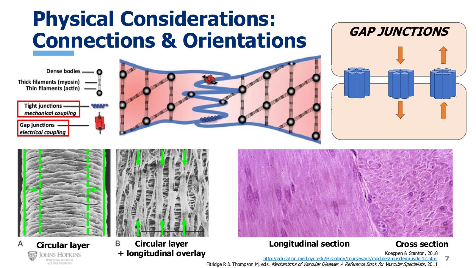
Slide 7. Circular and longitudinal layers, gap junctions, and the two viewing angles for smooth muscle tissue.
All smooth muscle cells are connected to their neighbors through membrane proteins that physically and mechanically couple them together. Tight junctions are pictured, though many types of mechanical coupling protein exist. You do not need the specific names, only that proteins tie adjacent cells together so a whole region contracts as one and transmits its contractile force through its neighbors.
Gap junctions connect cell membranes and allow electrical signals to pass directly from cell to cell with no additional neural stimulation. They work as essentially non-selective ion channels bridging the gap between membranes, joining the cytoplasm of one cell to the next. High intracellular concentrations of positive ions diffuse from cell to cell, depolarizing neighbors. Gap junctions are what let single-unit smooth muscle synchronize its depolarization.
Circular layer. Cells wrapping around the circumference of a cylindrical organ, as in a blood vessel. When they contract and shorten, the muscle tightens around the wall like cinching a belt, and the vessel narrows.
Longitudinal layer. Overlaid on a deeper circular layer, as in the intestinal wall. Contraction shortens the length of the organ. In the intestines the circular and longitudinal layers take turns, alternating between constriction and shortening. That coordinated action is peristalsis, which pushes food farther down the digestive tract.
Viewed from the side with a full view of cell length tip to tip, that is a longitudinal section. It lacks the crisp resolution skeletal muscle had, but a general visual grain runs top to bottom and nuclei are visible. Cut crosswise through the thickest part of the cells and that is a cross-section, where cells look like small circles packed closely together.