4B · Smooth Muscle Function
The general template
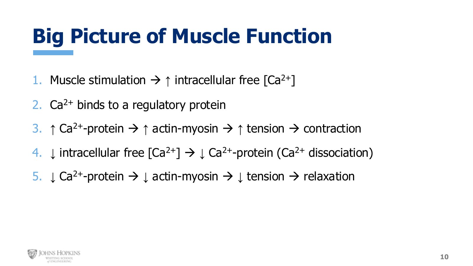
Slide 10. The five-step pattern that holds for every muscle tissue. Only the specifics change.
Before the details, the pattern that holds for every muscle tissue. Stripping out the skeletal specifics leaves a unifying way to view all muscle, and it places unfamiliar smooth muscle details into a broader context.
Muscle stimulation raises intracellular free calcium.
Calcium binds a calcium-sensitive regulatory protein.
More calcium bound to that protein means more actin-myosin interaction, more tension, contraction.
The stimulus goes away, intracellular free calcium falls, and calcium dissociates from the regulatory protein.
Less calcium bound means less actin-myosin interaction, less tension, relaxation.
Smooth muscle stimulation
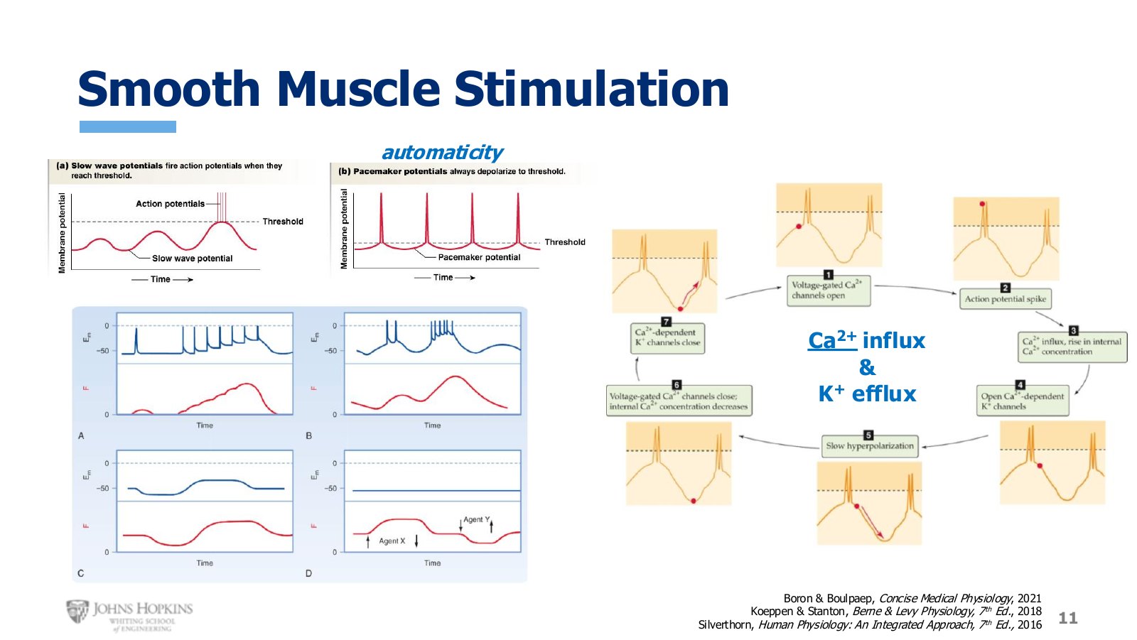
Slide 11. Four different membrane potential patterns, each producing its own force response. Blue is membrane potential, red is force.
Smooth muscle action potentials come in many waveforms. Slow wave potentials behave like graded potentials, and when membrane potential reaches threshold they produce an action potential spike.
The ions driving them differ from what we are used to. Potassium efflux still produces repolarization and hyperpolarization, but depolarization in both the graded and action potential waveforms appears to come from calcium influx, not sodium influx. That works because calcium carries a positive charge just like sodium, and its inward electrochemical gradient is very strong just like sodium’s, so calcium rushes in when voltage-gated calcium channels open.
Other patterns appear too:
Pacemaker potentials. Some smooth muscle cells spontaneously and regularly depolarize on their own, giving a repeating, reliable rate of self-generated depolarization. This is automaticity, and it spreads to all adjacent cells.
Summation of action potentials. Action potentials themselves undergo summation, and force production sums with them. Graded potential summation and force summation are familiar; summing the action potential waveform is new.
Subthreshold changes. Prolonged changes in membrane potential that never meet threshold still raise force when the membrane becomes less negative and lower it when the membrane becomes more negative.
No electrical change at all. Smooth muscle responds directly to chemical ligands, where one drug raises force and another lowers it with no change in membrane potential.
Physical stretch can also stimulate smooth muscle. Smooth muscle in one organ may be stimulated one way while smooth muscle in another organ is stimulated entirely differently. It is a flexible, versatile tissue that does not follow all the rules learned so far, and that is fine. Every route ends the same place: an increase in intracellular free calcium.
Two routes to raising calcium
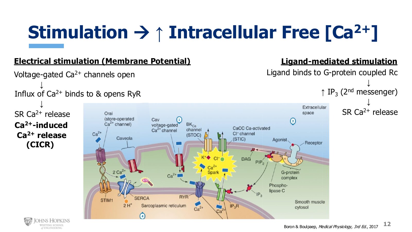
Slide 12. Left, the electrical route through voltage-gated calcium channels and CICR. Right, the ligand route through a G protein-coupled receptor and IP₃.
Electrical. The change in membrane potential travels across the sarcolemma and down into the caveolae, opening voltage-gated calcium channels and letting extracellular calcium flow in. That influx directly raises intracellular calcium, but it does something else too. The entering calcium ions attach to binding sites on the ryanodine receptor, which opens and releases stored calcium from the sarcoplasmic reticulum into the cytoplasm.
Here the ryanodine receptor acts as a ligand-gated ion channel, with calcium itself as the ligand. An influx of extracellular calcium stimulating a release of stored calcium is calcium-induced calcium release, or CICR.
Ligand-mediated. The agonist could be almost anything that stimulates the receptor: a neurotransmitter, a hormone, a chemical released from a nearby cell, or a drug. It binds a G protein-coupled receptor, which stimulates the enzyme phospholipase C. That enzyme takes one of the membrane phospholipids and breaks it into two pieces:
DAG, diacylglycerol, a hydrophobic second messenger that exerts its effects up at the membrane.
IP3, inositol triphosphate, a hydrophilic second messenger that enters the cytoplasm.
IP3 binds the IP3 receptor on the sarcoplasmic reticulum, also a ligand-gated ion channel, which opens and lets stored calcium flow into the cytoplasm.
Calcium binds calmodulin
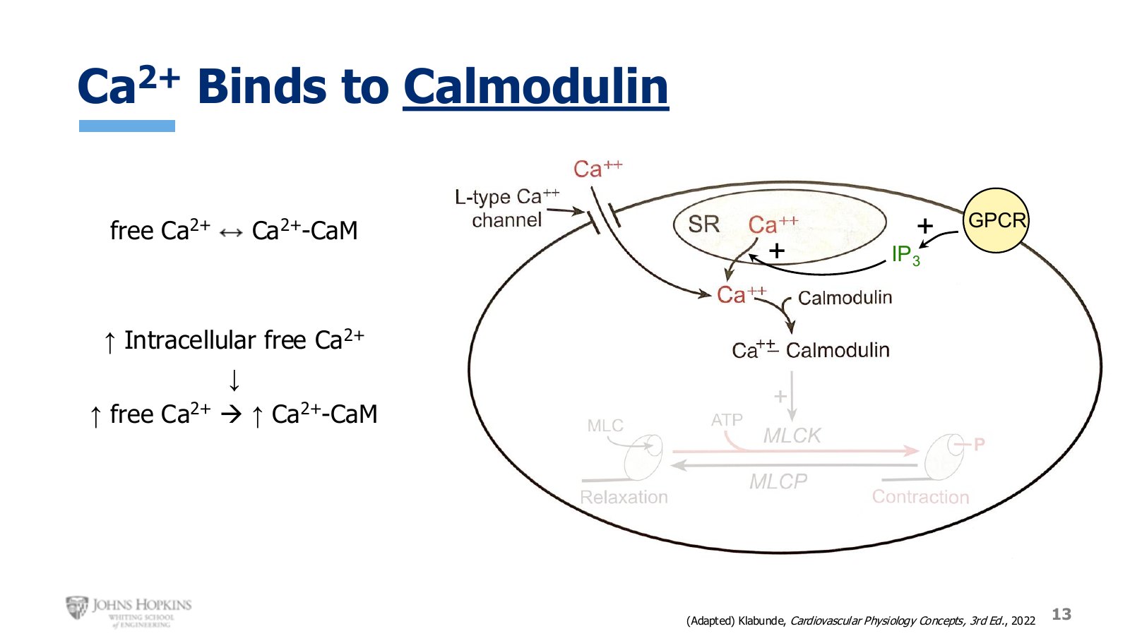
Slide 13. Calcium from both sources binds calmodulin. Le Chatelier again, at a new and higher equilibrium.
Smooth muscle has no troponin. Calcium from both extracellular and intracellular sources binds a regulatory protein called calmodulin, abbreviated CaM.
Le Chatelier’s principle applies exactly as it did with troponin. Before stimulation, free and bound calcium sit in equilibrium. The spike in intracellular free calcium disrupts it, the equation shifts to favor binding, and calcium-bound calmodulin rises.
The thick filament does the regulating
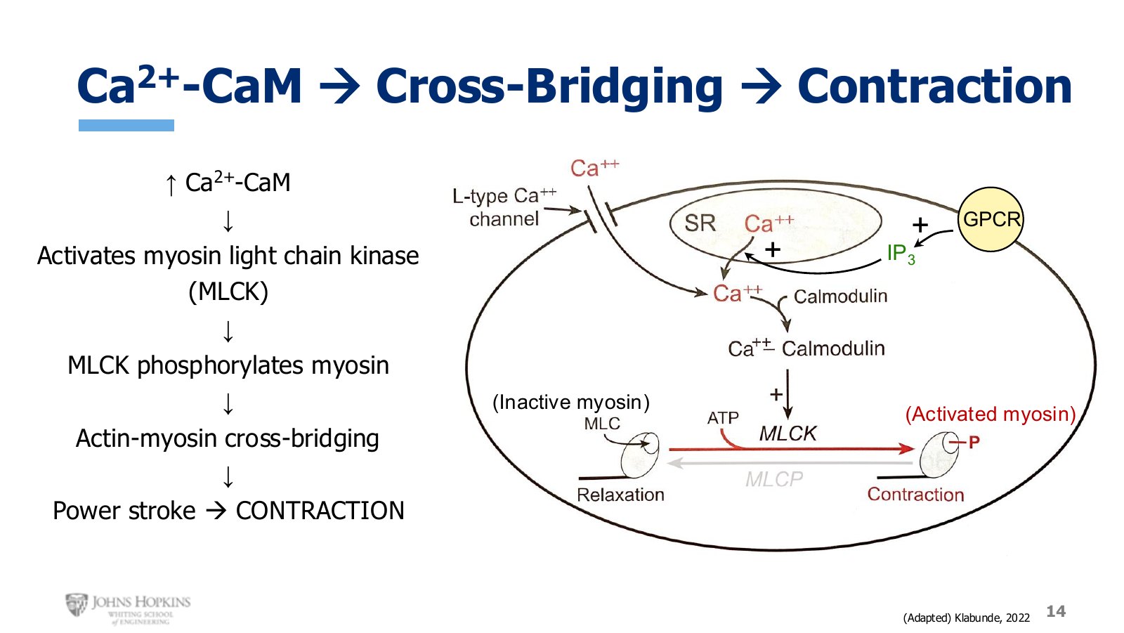
Slide 14. MLCK phosphorylates the myosin light chain. Phosphorylated myosin is the activated state.
This is the most distinctive difference between skeletal and smooth muscle. Regulation of contraction happened on the thin filament in skeletal muscle, where troponin and tropomyosin are. In smooth muscle it happens on the thick filament, and all the attention is on the myosin head.
Calcium binds calmodulin.
The calcium-calmodulin complex activates an enzyme, myosin light-chain kinase, or MLCK.
A kinase phosphorylates things, sticking a phosphate group onto a protein and changing how it functions. MLCK phosphorylates the light chain of myosin, part of the myosin head.
Phosphorylated myosin is the activated state, and that is what gives myosin permission to bind actin.
Actin-myosin cross-bridging proceeds through the sliding filament theory from Module 3, ending in a power stroke. Contraction.
Lowering calcium
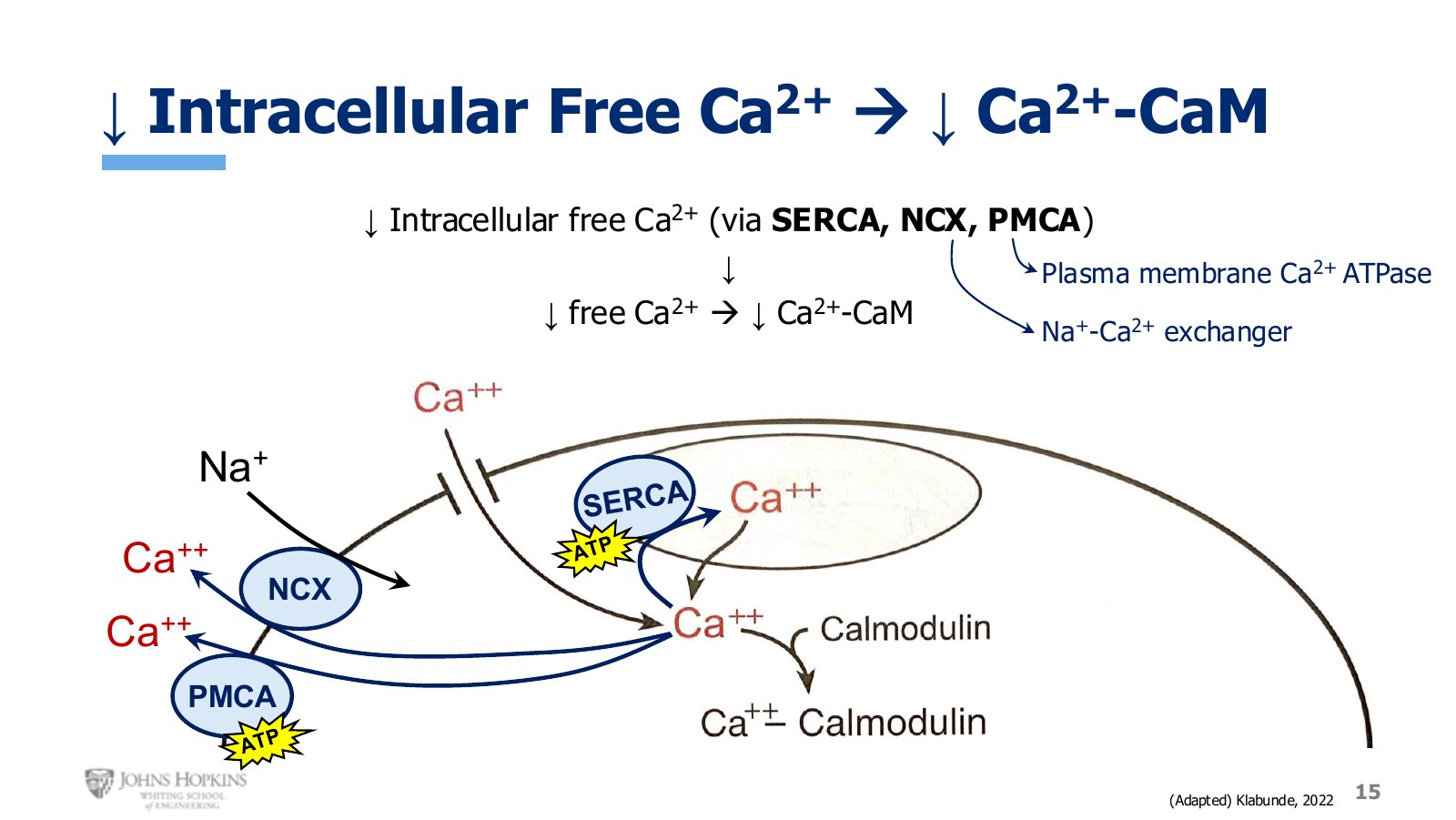
Slide 15. Three transport mechanisms clear calcium: SERCA into the SR, NCX and PMCA out of the cell.
Three calcium transport mechanisms bring free calcium down.
SERCA pump, familiar from skeletal muscle, pumping calcium back into the sarcoplasmic reticulum. Accounts for the majority of the decrease.
Sodium-calcium exchanger, or NCX, passively transporting calcium out of the cell as sodium flows down its gradient in. Second largest contribution.
Plasma membrane calcium ATPase, or PMCA, a calcium pump in the cell membrane actively pumping calcium out. Typically only a small contribution.
The big drop in free calcium disrupts equilibrium again, now favoring dissociation of calcium from calmodulin.
Relaxation
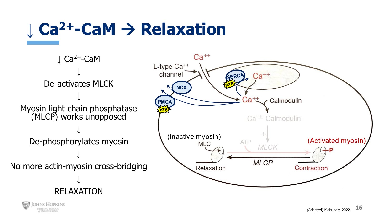
Slide 16. With MLCK deactivated, MLCP works unopposed and dephosphorylates myosin back to its inactive state.
Falling calcium-calmodulin deactivates myosin light-chain kinase, so myosin phosphorylation stops. But working in the background the whole time has been myosin light-chain phosphatase, or MLCP. With the kinase deactivated, the phosphatase works unopposed, giving net dephosphorylation of myosin.
Dephosphorylated myosin is the inactive state and cannot bind actin. No more cross-bridging, no more power strokes. That is relaxation in smooth muscle.
Contraction dynamics
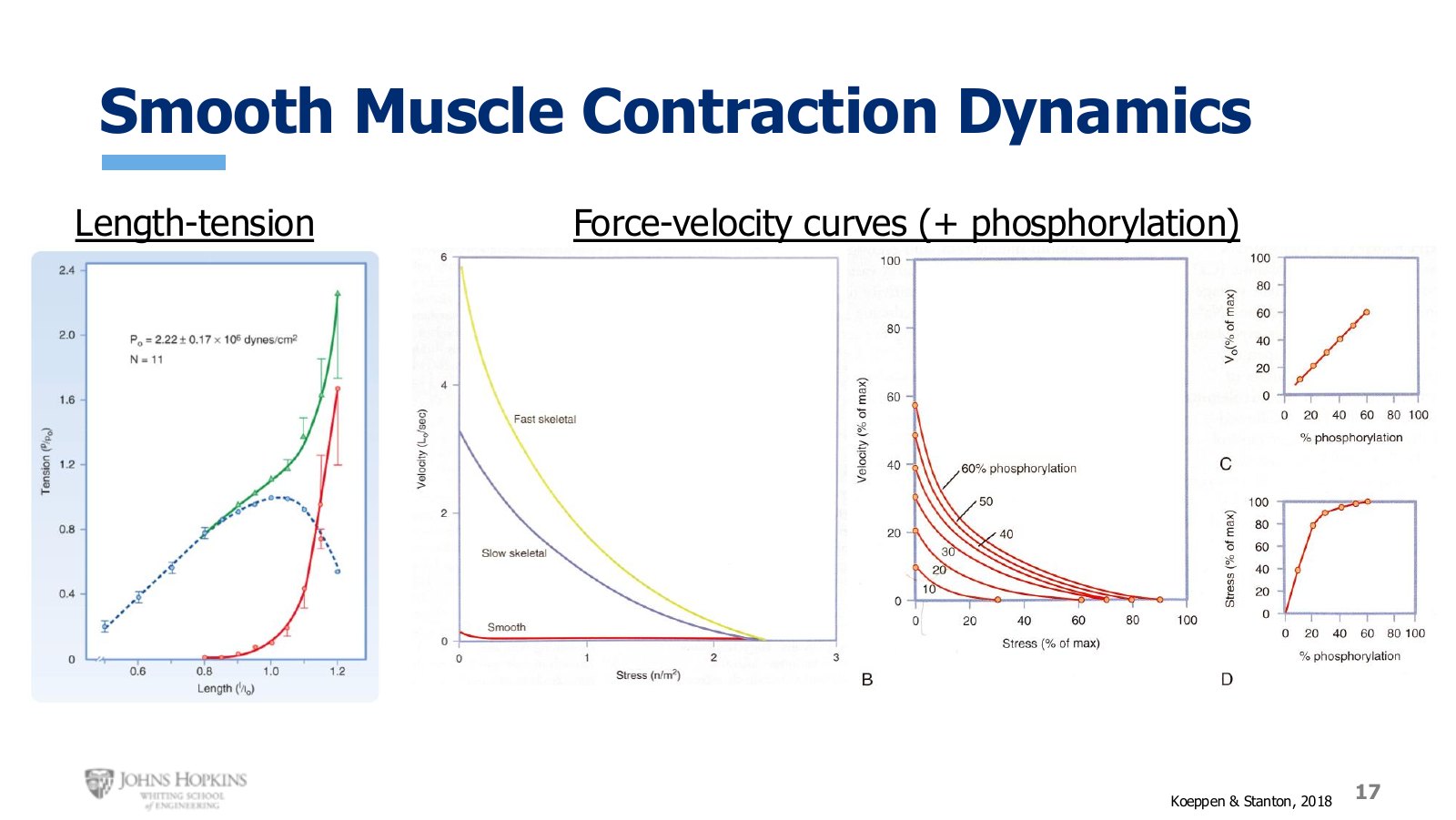
Slide 17. Length-tension on the left. On the right, a family of force-velocity curves set by how much myosin is phosphorylated.
Length-tension. Smooth muscle shows the same relationship skeletal muscle did, with the passive tension curve, the active tension curve, and the total tension curve. The one difference to keep in mind is that both axes are normalized, giving values relative to an optimal value of 1.0. Smooth muscle stretches more and shortens more than skeletal muscle, and can produce twitch contractions of very different tensions even at the same resting length. The normalized scales mask those differences.
Force-velocity. Roughly the same curve shape, with the fastest shortening velocity against zero load and velocity slowing as load increases. But the smooth muscle curve sits so far down the graph, its shortening velocity so slow most of the time, that nothing meaningful can be read off it without zooming in.
Zoomed in, a whole family of curves appears, set by how much myosin is phosphorylated at any moment. At 60% phosphorylated, a given load produces higher shortening velocity, and the muscle can contract against a much greater load before reaching an isometric state. At 10% phosphorylated, shortening velocity for the same load is much lower and the isometric point arrives at a much lower load.
That makes sense. More myosin phosphorylation means more actin-myosin cross-bridging and more power strokes, giving faster and stronger contractions.