4D · Introduction to Cardiac Muscle
Cardiac muscle is found only in the walls of the heart, helping the hollow chambers contract with enough pressure to propel blood out of the heart and through the blood vessels. It is also called the myocardium: myo- means muscle, cardio- means heart.
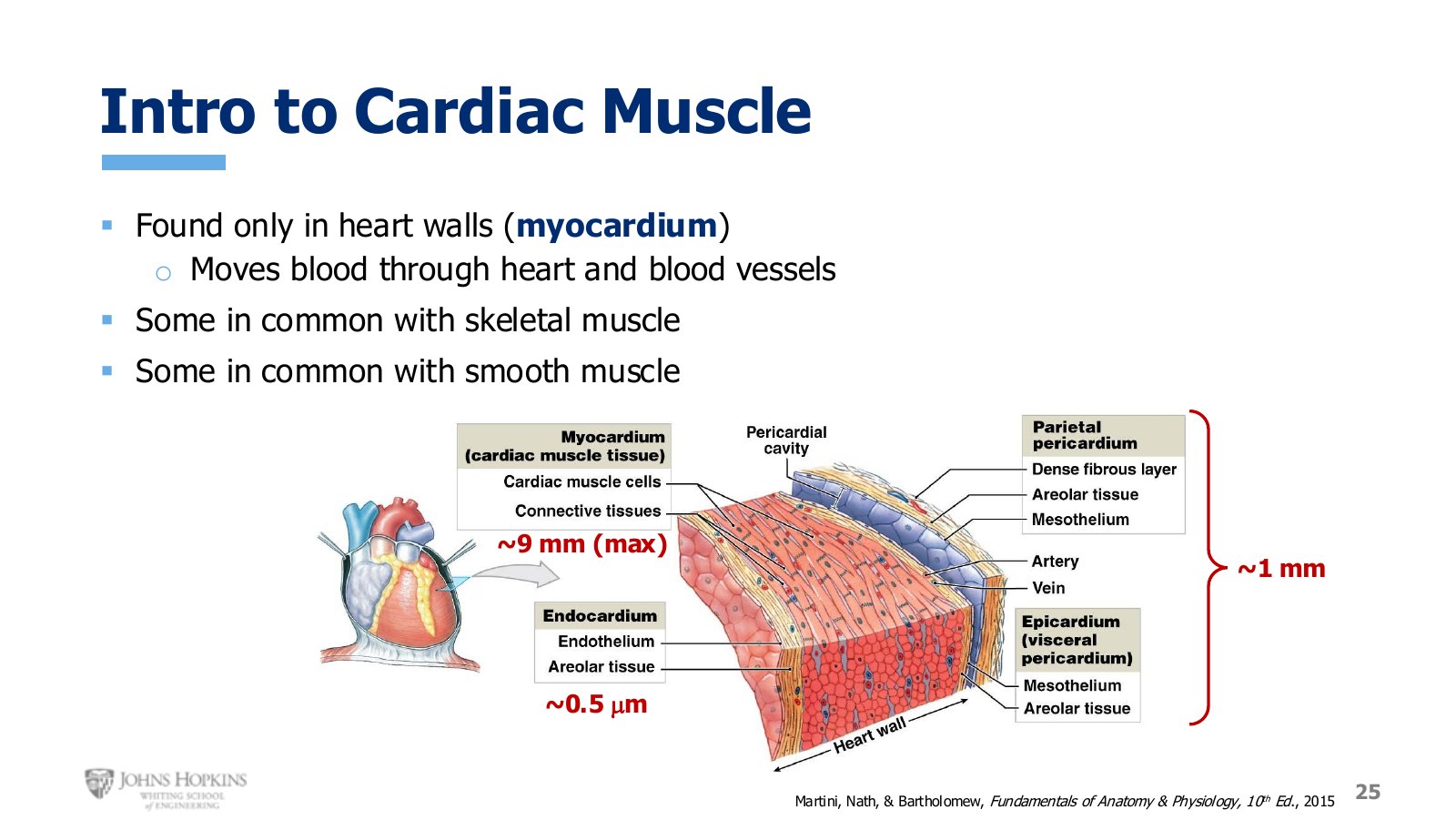
Slide 25. The layers of the heart wall, with the thick myocardium between two much thinner layers.
Layers of the heart wall
Layer | Thickness | What it does |
|---|---|---|
Pericardium | ~1 mm | Connective tissue layers on the outside. The pericardial cavity between them holds lubricating pericardial fluid, minimizing friction as the heart constantly changes shape and rubs against nearby organs. |
Myocardium | up to 9 mm | The thick meaty muscle in the middle. Varies with location in the heart. |
Endocardium | ~0.5 µm | Lines the hollow chambers. This is the tissue blood actually touches. |
On the slide the layer thicknesses are visually tweaked so they all fit on one image and stay visible. The real proportions are above.
Structure
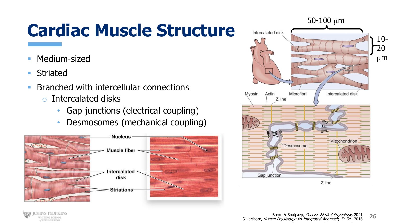
Slide 26. Branched, striated cells joined end to end at intercalated disks.
Cardiac muscle cells are medium-sized, 50 to 100 micrometers long and 10 to 20 micrometers thick. Much smaller than skeletal muscle cells, but larger, or at least thicker, than smooth muscle cells.
They are striated, because their contractile filaments are also organized into neatly repeating sarcomeres, though the striations are not as bold or thick as in skeletal muscle. They are clearer on the artist’s sketch than in the microscope image.
Cardiac cells branch, maximizing points of contact with surrounding cells. Where two membranes join, the junction appears darker under the microscope and is called an intercalated disk. Zoom in and it contains both mechanical coupling junctions called desmosomes and electrical coupling junctions called gap junctions, the same gap junctions seen in smooth muscle. Together they let cardiac muscle depolarize and then contract as a synchronous unit.
What it shares with skeletal muscle
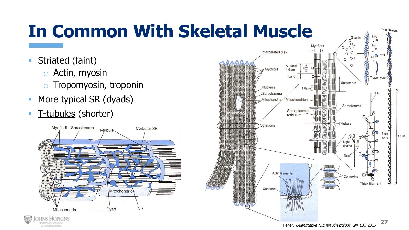
Slide 27. Sarcomeres, the full filament protein set, and T tubules — but a dyad rather than a triad.
Striated, meaning the same myofibril and sarcomere organization.
The same filament proteins: actin, myosin, tropomyosin, and troponin. Troponin is back.
T tubules, though shorter than in skeletal muscle.
A formal junction of sarcoplasmic reticulum with T tubule, but a dyad rather than a triad. In skeletal muscle two patches of sarcoplasmic reticulum converged on one point of the T tubule, three structures, a triad. In cardiac muscle one patch converges on the T tubule, two structural elements, a dyad.
What it shares with smooth muscle
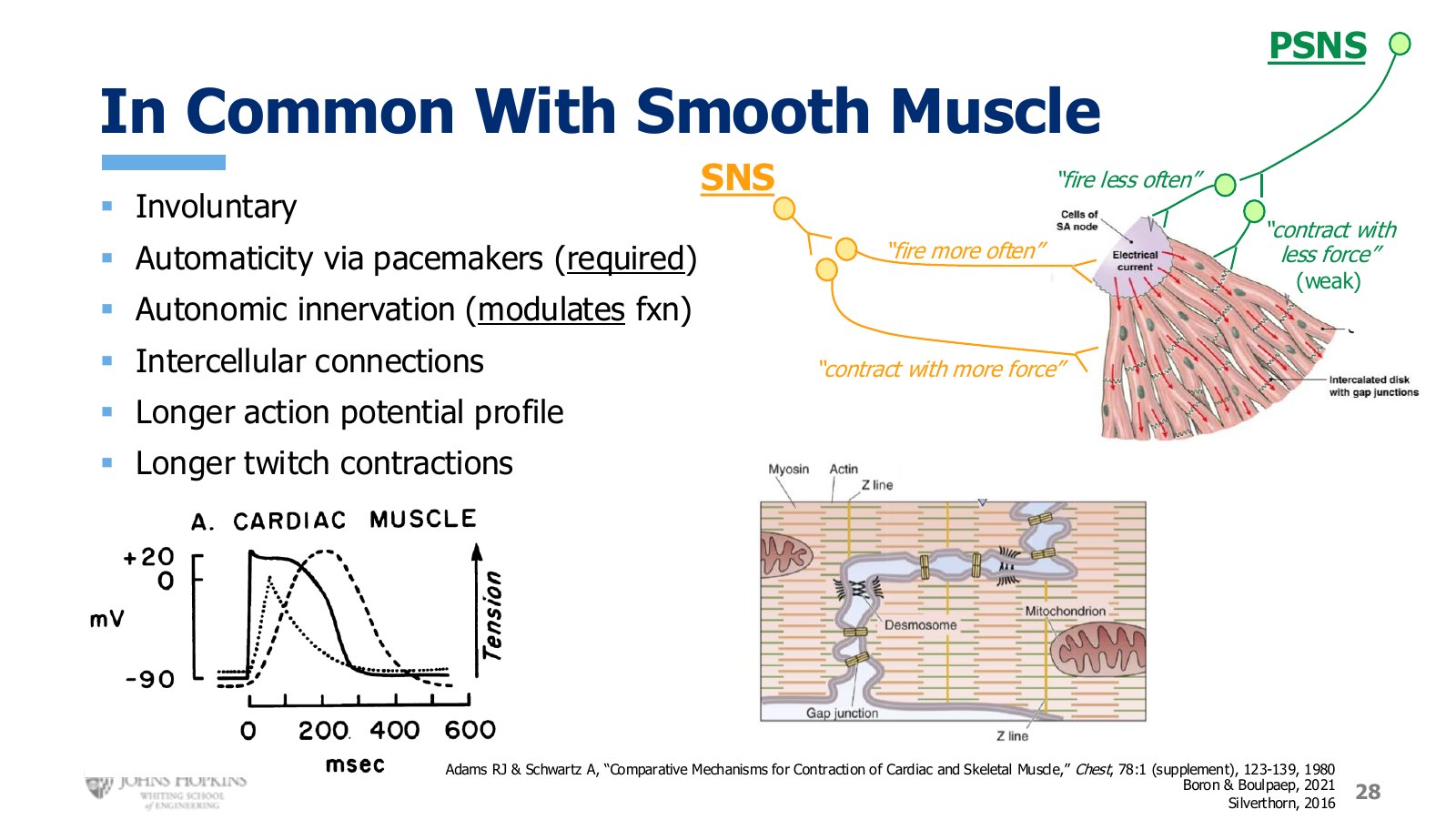
Slide 28. Autonomic nerves modulate rather than command. Sympathetic fires more often and contracts harder; parasympathetic does the reverse.
Involuntary. No conscious control over contraction or relaxation.
Automaticity. Regular spontaneous depolarizations arising within the muscle tissue itself.
Autonomic innervation, sympathetic and parasympathetic.
Intercellular connections, both physical and electrical.
Longer action potential profile and longer twitch contractions than skeletal muscle.
The autonomic nerves are also different here. They do not stimulate the heart to depolarize and contract, since the pacemaker cells bear that burden alone. The nerves modulate. Sympathetic nerves tell the pacemaker cells to fire more often than baseline, and tell the muscle cells to contract more forcefully than baseline. Parasympathetic nerves tell the pacemaker cells to fire less often, and the muscle cells to contract with less force, though that last effect is weak enough that it rarely comes up.
The action potential takes about 200 milliseconds, the twitch about 200 milliseconds to peak tension and another 200 to relax back to baseline. These long action potentials and long twitches are hallmarks of cardiac muscle, and the tracings return in later modules.